Pink Pulseless hand – Evaluation and Decision making: Is there a Consensus?

Vol 1 | Issue 1 | July-Sep 2015 | page:19-22 | Venkatadass K.


Authors : Venkatadass K[1].

[1] Consultant Paediatric Orthopaedic Surgeon, Ganga Medical Centre & Hospitals, Coimbatore, India.

Address of Correspondence
Dr. K. Venkatadass
Ganga Medical Centre & Hospitals, Coimbatore, India.
Email- venkatpedortho@gmail.com


Abstract

Background: The standard of care for the initial treatment of pulseless supracondylar fracture of the humerus is emergency closed reduction and percutaneous pin stabilization. Some of these patients remain pulseless even after closed reduction and pin fixation with a well perfused hand. The management of this so-called pink pulseless hand still remains controversial. The options described in the literature are either of the two extremes of just observation or exploration of the vessel and vascular repair if needed. There are no clear guidelines on when to explore a pink pulseless hand. This article reviews the current literature on this gray area with recommendations on the process of evaluation and decision-making in pink pulseless hand.
Keywords: Supracondylar humerus fracture, vascular injury, pink pulseless hand.


Introduction
The incidence of vascular injuries associated with displaced supracondylar fractures of humerus in children is about 10-20% [1,2,3]. It is reported to be more common in extension type fractures due to the close proximity of the proximal fragment to the neurovascular bundle [4,5]. In a child presenting with a pulseless supracondylar humerus fracture an urgent closed reduction with percutaneous pin stabilization is recommended. In majority of these patients the injured limb gets back the pulse and the hand appears well perfused after closed reduction and pin fixation. These patients are then treated as any other patient with supracondylar fracture humerus without any additional special precaution [3]. There is no confusion in the literature regarding the management of those patients in whom the limb remains pulseless, pale and unperfused after a closed reduction and stabilization [3]. Emergency exploration of the artery and arterial repair if needed to get back the circulation of the affected limb is the current recommendation. But there is still no consensus on the management of those limbs, which remain pulseless after closed reduction and pinning, but are pink and well perfused. The main reason for this confusion is lack of details of natural history of this entity [2]. The current literature on this enigmatous situation has been reviewed and a recommendation based on the available literature and our experience for the management of pink pulseless hand in supracondylar fracture humerus is presented.

Mechanism of vascular injury
The mechanism of injury to the neurovascular structures following supracondylar fracture of the humerus has been described in great detail by Meyerding as early as 1936 [6]. He was the first to study the configuration of the fracture in detail and propose that in extension type supracondylar fractures, the injuring force carries the distal fragment posteriorly stripping the posterior periosteum. The sharp anterior fragment pierces the anterior periosteum and brachialis and injures the neurovascular bundle, which lies in close proximity anteriorly. The vascular insult could either be due to compression from the fragment, spasm or thrombosis or rarely complete arterial transection. Most times the absence of pulse might just be due to the compression of grossly displaced fragments. A gentle closed reduction would relieve the compression and artery becomes pulsatile again. Louahem et al. [7 described 26 patients with a pink pulseless hand in a series of 210 patients with severely displaced supracondylar fractures. In 21 cases, the pulses returned immediately after closed reduction of the fracture.

Collateral circulation
The exuberant collateral circulation around the elbow has been credited with maintaining the vascularity of the limb in patients managed without vascular exploration [8]. The radial recurrent artery arises distal to the elbow and anastomoses with the radial collateral branch of the profunda brachii. The superior ulnar collateral artery is the other main descending collateral. It arises from the brachial artery, a little below the middle of the arm and anastomoses with the posterior ulnar recurrent and inferior ulnar collateral arteries [8].

What is pink pulseless hand?
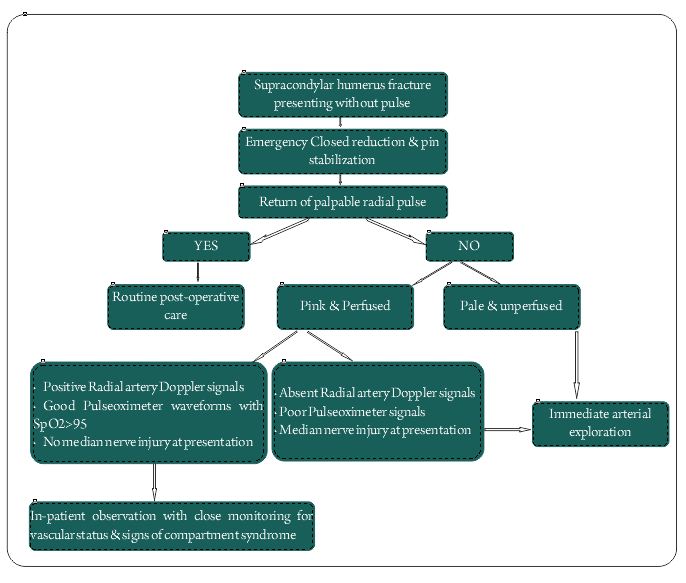
There is variable use of this terminology in the literature. Some authors use this terminology to label those supracondylar fractures presenting without a pulse with a well perfused hand. While there is no controversy regarding the management of these fractures, more than 50% of these pink pulseless limbs would turn pulsatile just after closed reduction. The limb appearing pink and perfused on presentation mainly depends on the time duration since injury, as it needs some time for the collateral circulation to be established. None of the authors had looked into the time since injury and its correlation to perfusion status at presentation. Ideally, a pink pulseless limb is one that remains pink and well perfused without a palpable pulse following closed reduction and pinning of a pulseless supracondylar fracture [2]. These are the ones that pose management controversy as to whether it needs urgent exploration for the vessel or a closed monitoring for the vascular status and just observation. Though there is some evidence in the literature supporting immediate exploration, more recent evidence seems to be in favour of in-patient observation and close monitoring [3].

Role of Doppler
There has been increased interest in the role of colour Doppler in the process of evaluation and decision making for pink pulseless hands. White et al has recommended the use of colour Doppler to assess the severity of arterial injury following closed reduction and pinning to decide on further management. A colour Doppler evaluation of the brachial artery would help to differentiate between spasm, thrombosis and complete transection. It is important to remember the fact that there is no question of brachial artery injury in this scenario and the real issue is about the adequacy of the collateral circulation to maintain the viability and function of hand. Doppler evaluation of the brachial artery might infact increase the number of explorations of the artery. Valentini et al has reported the use of color-coded duplex scanning (CCDS) and ultrasound velocimetry (UV) of the hand as an additional tool for evaluation in all their patients with pink pulseless hands [9]. In their series, all seven patients with pink pulseless hand were found to have brachial artery injury by Doppler and all of them were treated by arterial repair. But, there are no clear guidelines on severity of arterial injuries on colour dopper and their management.  Using Doppler to assess the radial artery in a pink pulseless hand helps to assess the adequacy of the collateral circulation. Weller et al [10] in their series of 54 pulseless supracondylar humerus fractures have documented that 26 patients regained the pulse after closed reduction, 20 remained pulseless after closed reduction but radial artery Doppler signals were picking up and 4 others had absent pulse as well as Doppler signals. All four were taken up immediate surgical exploration and found to have arterial injury requiring repair. All 20 patients who had pulse detected by Doppler but had no palpable radial pulse were observed. One of the 20 developed late ischaemia after nine hours and was taken up for surgical exploration. Shah et al have included triphasic radial artery doppler signal in their algorithm for decision making in pink pulseless limbs and recommends immediate surgical exploration for patients who do not have triphasic radial artery doppler signals.

Figure

Proponents for Immediate Exploration
White et al [11] after a systematic analysis of pink pulseless supracondylar fractures have concluded that there is significant arterial injury in 70% of patients and thus vascular exploration may limit the chances of late complications in these patients. They have also stated that with reported patency rates of more than 90% it is worthwhile considering exploration and arterial repair in these patients.  Korompilias et al. [7] reported on five patients with a pink pulseless hand and recommended vascular exploration for the restoration of brachial artery patency, even in the presence of a viable well-perfused hand after an attempt at closed reduction. Copley et al [13] in their series of 17 patients with pulseless supracondylar fractures had a return of pulse in 14 of them following closed reduction. All the three patients were taken up for exploration and 2 of the fourteen patients who developed loss of pulse over the next 24 hours were also explored. They recommend immediate exploration if pulse is absent after closed reduction as a measure towards prevention of late complications.  Blakey et al. found that twenty three of twenty-six patients with a pink, pulseless hand following initial management had some evidence of ischemic contracture, and they advocated for urgent exploration when the pulse does not immediately return after closed reduction [14]. Mangat et al [15] reported on the predictive value of co-existing median or anterior interiosseous nerve injury after studying a series of patients with nerve injury who underwent exploration. A significant relationship was found between preoperative median and anterior interosseous nerve deficits and vascular entrapment and tethering of the nerve at the fracture site. The authors recommended early exploration for patients with a Gartland type-III supracondylar fracture when they have coexisting anterior interosseous or median nerve palsy as the benefits of exploration outweigh the disadvantages.  In a recent study by Scannell et al [17], the authors have tried to correlate the presence of median or anterior interosseous nerve with patency of brachial artery at long term follow-up in 20 twenty patients. In their series, median nerve palsy had good prediction of brachial artery occlusion while anterior interosseous nerve palsy did not predict brachial artery occlusion.

Proponents for Observation
Many authors are in favour of observation and close monitoring of the vascular status for pink pulseless hands. In contrast to the general belief that the literature in vascular surgery would be more in favour of arterial exploration, the recent papers in vascular surgery are recommending observation in case of pink pulseless hands [3].  Choi et al [18] presented the largest series of 33 patients with pink pulseless hands and concluded that in patients presenting with well perfused hand, fracture reduction and pinning alone would be sufficient treatment. Scannell et al [17] have reported the long term results of their series of 20 patients of pink pulseless hand that were treated by observation. All 20 had good functional outcome except one who had chondrolysis of the distal humerus. They also recommended long term follow-up of these patients for radiographic evidence of osteonecrosis as three of their 20 patients with pink pulseless hands developed avascular necrosis of the trochlea. Weller et al [10] in their analysis of 20 patients with pink pulseless hands have concluded that lack of palpable radial pulse is not an absolute indication for arterial exploration if Doppler signals and capillary refill is good suggesting a well perfused hand. Matuszewski [19] has published his follow-up results of pulseless supracondylar humerus fractures have concluded that children who, after satisfactory closed reduction, have a well-perfused hand but absent radial pulse do not necessarily require routine exploration of the brachial artery. Sabharwal et al [20], in their follow-up study patients with pulseless supracondylar fracture who were treated with arterial exploration and revascularisation found a high rate of asymptomatic reocclusion and residual stenosis and hence opined that collateral circulation would have been adequate to maintain a viable extremity. Garbuz et al [21] in 1996 presented the outcome of treatment of supracondylar fractures with absent radial pulse from the Hospital for Sick Children, Toronto. In their series of 22 patients, five had pink perfused pulseless hands who were managed by close observation and all had excellent functional outcomes. They concluded that absent pulse is not an absolute indication for exploration, provided the hand remains well perfused and compartment syndrome does not develop.

Is there a Consensus?
Though the literature is filled with publications on pink pulseless hand in supracondylar fractures, there still seems to be no consensus on the management of this condition. The AAOS guidelines [22] for the management of supracondylar fractures of humerus in children published in 2010 stated that ”We cannot recommend for or against open exploration of the antecubital fossa in patients with absent wrist pulses but with a perfused hand after reduction of displaced pediatric supracondylar humerus fractures” as there was no strong evidence supporting either observation or exploration. Five years down the line, the question still remains unanswered as far as evidence goes. But, what has changed over the years is that now we have more objective ways of assessing the perfusion rather than just relying on pink colour of the hand and capillary refill. The use of Doppler ultrasound and pulseoximeter signals to assess the perfusion of limb have come into vogue [23]. The presence of associated median nerve injury is more predictive of a significant arterial injury and hence these patients should be considered for exploration.
Hence in the present scenario, three factors needs to be considered in the decision making process of pink pulseless hand:
1.Presence of radial artery Doppler signals
2.Presence of good pulseoximeter waveforms and oxygen saturation >95%.
3.Intact Median Nerve function.

If all the three criteria are met, the recommendation is to observe the child closely for circulation and symptoms of compartment syndrome. If all three are absent, it is an indication of poor perfusion and it is an indication for arterial exploration. The combination of absence of radial artery Doppler signals and absence of pulse oximeter signals again indicates poor perfusion and favors exploration.  There is no evidence to comment on other scenarios of either isolated median nerve palsy or isolated absence of radial artery Doppler signals or pulse oximeter signals and their combinations. There are no studies, which have documented all these factors for all their patients, and we are not sure whether a limb can have absent radial artery Doppler signals with good pulse oximeter waveforms. These would rather be hypothetical situations and if someone comes across such a situation in clinical practice, the best would be to individually assess the case and decide for exploration versus observation. However, considering the complications and the reported incidence of significant arterial injuries up to 70% in patients without a positive radial artery Doppler signal, it may be a safer option to consider exploration in these patients. The use of Doppler of the brachial artery to know the severity of the arterial injury and taking it as a sole factor for considering exploration is not justifiable, as the limb can still have good collateral circulation.  Thus in conclusion both clinical and diagnostic methods have to be taken into account while making a balanced decision in terms on observation or surgical exploration of a pink pulseless hand [3,24].


References

1. Schoenecker PL, Delgado E, Rotman M, Sicard GA, Capelli AM. Pulseless arm in association with totally displaced supracondylar fracture. J Orthop Trauma 1996; 10:410–415.
2. Robb JE. The pink, pulseless hand after supracondylar fracture of the humerus in children. J Bone Joint Surg Br. 2009 Nov;91(11):1410-2.
3. Badkoobehi H, Choi PD, Bae DS, Skaggs DL. Management of the pulseless pediatric supracondylar humeral fracture. J Bone Joint Surg Am. 2015 Jun 3;97(11):937-43.
4. Korompilias AV, Lykissas MG, Mitsionis GI, Kontogeorgakos VA, Manoudis G, Beris AE (2009) Treatment of pink pulseless hand following supracondylar fractures of the humerus in children. Int Orthop 33(1):237–241.
5. Matuszewski Ł. Evaluation and management of pulseless pink/pale hand syndrome coexisting with supracondylar fractures of the humerus in children. Eur J Orthop Surg Traumatol. 2014 Dec;24(8):1401-6.
6. MEYERDING HW. Volkmann’s ischemic contracture associated with supracondylar fracture of humerus. Journal of the American Medical Association, 1936:106:1139-1144.
7. Louahem DM, Nebunescu A, Canavese F, Dimeglio A. Neurovascular complications and severe displacement in supracondylar humerus fractures in children: defensive or offensive
strategy? J Pediatr Orthop B 2006; 15(1):51–57
8. Ramesh P, Avadhani A, Shetty AP, Dheenadhayalan J, Rajasekaran S. Management of acute ‘pink pulseless’ hand in pediatric supracondylar fractures of the humerus. J Pediatr Orthop B. 2011 May;20(3):124-8.
9. Benedetti Valentini M, Farsetti P, Martinelli O, Laurito A, Ippolito E. The value of ultrasonic diagnosis in the management of vascular complications of supracondylar fractures of the humerus in children. Bone Joint J. 2013 May;95-B(5):694-8.
10. Weller A, Garg S, Larson AN, Fletcher ND, Schiller JR, Kwon M, Copley LA, Browne R, Ho CA. Management of the pediatric pulseless supracondylar humeral fracture: is vascular exploration necessary? J Bone Joint Surg Am. 2013 Nov 6;95(21):1906-12.
11. White L, Mehlman CT, Crawford AH. Perfused, pulseless, and puzzling: a systematic review of vascular injuries in pediatric supracondylar humerus fractures and results of a POSNA questionnaire. J Pediatr Orthop. 2010 Jun;30(4):328-35.
12. Korompilias AV, Lykissas MG, Mitsionis GI, Kontogeorgakos VA, Manoudis G, Beris AE. Treatment of pink pulseless hand following supracondylar fractures of the humerus in children. Int Orthop. 2009 Feb;33(1):237-41.
13. Copley LA, Dormans JP, Davidson RS. Vascular injuries and their sequelae in pediatric supracondylar humeral fractures: toward a goal of prevention. J Pediatr Orthop. 1996 Jan-Feb;16(1):99-103.
14. Blakey CM, Biant LC, Birch R. Ischaemia and the pink, pulseless hand complicating supracondylar fractures of the humerus in childhood: long-term follow-up. J Bone Joint Surg Br. 2009 Nov;91(11):1487-92.
15. Copley LA, Dormans JP, Davidson RS. Vascular injuries and their sequelae in pediatric supracondylar humeral fractures: toward a goal of prevention. J Pediatr Orthop. 1996 Jan-Feb;16(1):99-103.
16. Luria S, Sucar A, Eylon S, Pinchas-Mizrachi R, Berlatzky Y, Anner H, Liebergall M, Porat S. Vascular complications of supracondylar humeral fractures in children. J Pediatr Orthop B. 2007 Mar;16(2):133-43
17. Scannell BP, Jackson JB 3rd, Bray C, Roush TS, Brighton BK, Frick SL. The perfused, pulseless supracondylar humeral fracture: intermediate-term follow-up of vascular status and function. J Bone Joint Surg Am. 2013 Nov 6;95(21):1913-9.
18. Choi PD, Melikian R, Skaggs DL. Risk factors for vascular repair and compartment syndrome in the pulseless supracondylar humerus fracture in children. J Pediatr Orthop. 2010 Jan-Feb;30(1):50-6.
19. Matuszewski Ł. Evaluation and management of pulseless pink/pale hand syndrome coexisting with supracondylar fractures of the humerus in children. Eur J Orthop Surg Traumatol. 2014 Dec;24(8):1401-6.
20. Sabharwal S, Tredwell SJ, Beauchamp RD, Mackenzie WG, Jakubec DM, Cairns R, LeBlanc JG. Management of pulseless pink hand in pediatric supracondylar fractures of humerus. J Pediatr Orthop. 1997 May-Jun;17(3):303-10.
21. Garbuz DS, Leitch K, Wright JG. The treatment of supracondylar fractures in children with an absent radial pulse. J Pediatr Orthop. 1996 Sep-Oct;16(5):594-6.
22. The treatment of pediatric supracondylar humerus fractures. AAOS Clinical Practice Guidelines Unit v1.0_092311. Summary of Recommendations. available from
http://www.aaos.org/research/guidelines/SupracondylarFracture/SupConFullGuideline.pdf
23. Soh RC, Tawng DK, Mahadev A. Pulse oximetry for the diagnosis and prediction for surgical exploration in the pulseless perfused hand as a result of supracondylar fractures of the distal humerus. Clin Orthop Surg. 2013 Mar;5(1):74-81.
24. Shah AS, Waters PM, Bae DS. Treatment of the “pink pulseless hand” in pediatric supracondylar humerus fractures. J Hand Surg Am. 2013 Jul;38(7):1399-403
.


How to Cite this Article: Venkatadass K. Pink Pulseless hand – evaluation and decision making: Is there a consensus?. International Journal of Paediatric Orthopaedics July-Sep 2015;1(1):19-22.         

Photo


(Abstract)      (Full Text HTML)      (Download PDF)


Supracondylar Humerus Fractures in Children: Epidemiology and Changing Trends of Presentation

Vol 1 | Issue 1 | July-Sep 2015 | page:3-5 | Sandeep V Vaidya.


Authors : Sandeep V Vaidya[1,2,3*].

[1] Children’s Orthopaedic Clinic, Thane.
[2] B J Wadia Children’s Hospital, Parel Mumbai
[3] Jupiter Hospital, Thane, Maharashtra, India

Address of Correspondence
Dr Sandeep V Vaidya
Director, Children’s Orthopaedic Clinic, Thane. India.
Email: drsvvaidya@gmail.com


Abstract

Diseases show a tendency to vary according to changing socio-economic trends and fractures too have shown this tendency. Paediatric supracondylar humerus fractures are one of the most common fractures seen by paediatric orthopaedic surgeons. There are few notable trends that have been reported and few other that I have personally noted in my practice and in practice of my colleagues. This article put together the changes reported in literature and tries to combine it with clinically relevant practical situations. Special focus is on fracture presentation and on decision making in management.
Keywords: Supracondylar Humerus fracture, classification, management.


Introduction:
Supracondylar humerus fractures in children are commonly seen in day to day practice. In this section, we study the epidemiology and changing trends of these fractures with respect to incidence, patient profile, types, modes of injury, treatment trends and complications.

Incidence:
Supracondylar humerus fractures (SHF) comprise 17% of all pediatric fractures and are second in frequency to forearm fractures. According to an epidemiological study, the incidence of fracture supracondylar humerus is 308/100000 per year in the general population. It is also the commonest pediatric fracture around the elbow. One epidemiological study identified supracondylar fractures in 206 out of 355 elbow fractures (58%) [1]. Barr reported a higher incidence of supracondylar humerus fractures during the vacations [2].

Age and sex:
If age distribution is considered, in the 0 to 7 year age group, SHF is easily the commonest fracture seen (28%) [3]. The mean age at which fracture supracondylar humerus occurs is 5 to 8 years [1,2]. Wilkins proposed that when a child falls on extended upper extremity, the patients who demonstrate hyperextension (cubitus recurvatum) of the elbow are more predisposed to have supracondylar fractures. The children who do not have hyperextension of the elbow tend to sustain fractures of the radius and the ulna, usually at the distal portion. Since ligamentous laxity with elbow recurvatum is seen in younger children, this explains the higher incidence of supracondylar humerus fractures in younger children and higher incidence of radius ulna fractures in older children.
Recently, there seems be increase in incidence of SHF in lower age group(less than 2 years). Fractures occurring in these very young children may pose a diagnostic dilemma because in many of these cases, the fracture line is extremely low and on plain radiographs may mimic a fracture lateral condyle humerus due to the largely cartilaginous component of the distal fragment. In such cases, additional imaging like MRI or arthrogram may be needed to differentiate these low supracondylar fractures from the lateral condyle fractures (Fig. 1). Another peculiarity of the low supracondylar humerus fractures is that such fractures can be complicated by Avascular necrosis of the trochlea with subsequent later sequelae.

Fig 1

Figure 1: (Case Courtesy Dr Sandeep Patwardhan)1a: Elbow radiograph`of a 2 year old child with fall on outstretched hand. The fracture line is extremely distal and only a flake of metaphysis is seen.1b,c: The fracture was treated by closed reduction and K wire pinning .

In most of the earlier studies, the fracture occurred much more commonly in boys than in girls. However in most of the recent series, the frequencies in girls and boys seems to be equalizing. Some series have actually reported a higher incidence in girls than boys[1,2]. This changing sex distribution may be attributed to more active participation of girls in sports activities.

Mode of injury:
The cause of fracture supracondylar humerus is accidental fall while playing in most of the cases (60 to 80 %). Road traffic accidents account for 10 to 20% of SHF [2]. High velocity trauma can lead to fractures with metaphyseal comminution or in rare cases fractures with intercondylar extension.
Child abuse is an uncommon etiology of SHF[4]. However Strait and colleagues reported supracondylar fractures from abuse in three of 10 abused children under the age of 3, and cautioned that SHF should not be assumed to have non-abusive causes without careful consideration [5].

Types:
Extension type is the commonest type, flexion type is seen in 1 to 3% cases [6]. The patients in the flexion-type group (mean age, 7.5 years) are significantly older than those in the extension-type group (mean age, 5.8 years). The fractures in flexion-type group are also more probable to require open reduction (31%) than those in the extension-type group (10%). The flexion-type group had a significantly increased incidence rate of ulnar nerve symptoms (19% vs 3% in the extension-type group) and need for ulnar nerve decompression [7].
Gartland classification is the commonest classification system used to grade supracondylar humerus fracture. Grade 1 fractures are the commonest, followed by Grade 2 and then Grade 3 [1,2].
In addition to these 3 types, Leitch et al described a type 4 fracture with multidirectional instability (unstable in both flexion and extension). This fracture type was noted in 9 out of 297 displaced fractures. These fractures are associated with high velocity trauma, the periosteal sleeve is completely torn and special manoeuvres are needed for closed reduction- pinning [8].
In extension type fractures the distal fragment may be displaced posteromedially or posterolaterally. Posteromedial displacement is commoner and seen in approximately 75% cases in most series. Posteromedial displacement of the distal fragment places the radial nerve at risk, whereas in fractures with posterolateral displacement the brachial artery and median nerve are at risk [9]. Bahk et al additionally classified extension type supracondylar fractures based on orientation of the fracture line in coronal as well as sagittal planes. In coronal plane, transverse fractures were the commonest (49%) followed by lateral oblique fractures (44%). Medial oblique (4%) and high transverse fractures (3%) were less common. Whereas transverse and lateral oblique fractures are amenable to lateral only pinning, the medial oblique and transverse fractures need to be fixed with medial-lateral cross pins [10].
High SHF are also being increasingly reported recently. Sen et al reported an incidence of high metaphyseal- diaphyseal supracondylar humerus fractures in 6 out of 182 fractures [11].

Treatment:
Blount in 1955 had cautioned against operative treatment in SHF citing the high incidence of complications following operative treatment [12]. However with significant advances in operative techniques and intraoperative imaging, operative treatment with Closed Reduction Percutaneous Pinning (CRPP) is easily the treatment of choice for displaced supracondylar humerus fractures [13]. Approximately 40% of SHF are treated operatively making it the commonest pediatric fracture to undergo operative treatment [2]. Cheng et al in an epidemiological study of 6493 fractures reported that the closed-reduction and percutaneous pinning rates for supracondylar humerus fractures increased 4.3 to 40% over a 10 year period from 1985 to 1995. The changes in treatment pattern were also accompanied by a corresponding decrease in the open-reduction rate and hospital stay periods from <10% to 38% of patients being discharged within 1 day of admission in the 10-year period [3].

The incidence of operative treatment is 0% in Grade 1 fractures, almost 50% for Grade 2 fractures, 100% for Grade 3 fractures and 100% for flexion type fractures. The incidence of open reduction is highest in flexion type fractures (50%) [2]. In an epidemiological study, out of 3235 children with displaced SHF treated operatively at a tertiary care children’s hospital at Toronto, 78.7% underwent operative treatment in the form of Closed Reduction Percutaneous Pinning (CRPP) whereas the remainder 21.8% underwent Open Reduction Internal Fixation (OR). There was a significant difference in the delay to surgery between the CRPP and OR groups [14]. In developed countries, there is a trend for more number of SHF are being treated by pediatric orthopaedic subspecialists. In New England, only 37% of SHF were treated by Pediatric Orthopaedic surgeons in 1991, this number rose to 68% in 1999. Kasser et al reported that in fractures treated by pediatric orthopaedic surgeons the length of hospitalization was lesser (1.4 ± 0.4 days) than for fractures treated by general orthopaedic surgeons (2.2 ± 0.6 days) [15]

Pin configurations, changing trends:
Pin configurations used by surgeons have shown a changing trend over the past decade. Several biomechanical studies published before 2005 revealed that crossed medial- lateral pin configurations are biomechanically stronger than lateral only pin configurations. Hence crossed medial- lateral pinning was preferred. However a major danger of the medial pin was iatrogenic ulnar nerve injury. Incidence of iatrogenic ulnar nerve injury with crossed medial- lateral pinning in various series has ranged from 0% to 6% [16,17]. Lyons et al reported iatrogenic ulnar nerve palsy in 19 out of 375 crossed medial- lateral pinning. 15 out of these 19 palsies recovered within 4 months after medial pin removal. However 4 palsies failed to recover, underwent ulnar nerve exploration and neurolysis [17]. A systematic pooled analysis of 32 trials comprising 2639 children suggests that there is an iatrogenic ulnar nerve injury for every 28 patients treated with the crossed pinning compared with the lateral pinning [16].
An inherent fallacy of the early biomechanical studies was that these studies were based on in-vitro findings wherein loads applied to create displacement were significantly higher than those which would be applied in-vivo wherein the fixation would be additionally supplemented with plaster slab application. Lee et al in their series of 61 consecutive lateral only pinning reported a zero incidence of loss of reduction as well as iatrogenic ulnar nerve palsy [18]. A randomized controlled study published in 2007 concluded that lateral entry only pinning did not result in increase incidence of loss of reduction as compared to crossed medial-lateral pinning [19]. A survey involving eight surgeons conducted in 2012 confirmed that this RCT had a significant influence on the surgeons’ practice. Five out of eight surgeons individually had a statistically significant change in their practice pattern for pin configuration. Except for certain selected fracture patterns, lateral only pinning is being increasingly used as the standard pin configuration for supracondylar humerus fractures [20].

Complications:
Complications of fracture supracondylar humerus include compartment syndrome, vascular injury, nerve injury (fracture related or iatrogenic) and malunion with cubitus varus deformity. The incidence of compartment syndrome is approximately 0.1% to 0.3% of all supracondylar humerus fractures [21]. Ipsilateral forearm fracture significantly increases risk of compartment syndrome [22]. In a study, the incidence of compartment syndrome was — % in fractures reduced and fixed within – hours of injury as compared to — % in fractures fixed after a delay of – hours.
The incidence of vascular injuries is approximately 20% and majority are associated with Grade 3 fractures [1, 23, 2]. Fractures with posterolateral displacement are more at risk for vascular injuries (approximately 65%) than fractures with posteromedial displacement (approximately 53%) [23]. If the hand is well perfused but pulseless, the great majority of the time fracture reduction is sufficient treatment. In contrast, patients presenting with a pulseless and poorly perfused hand have a nearly 50% chance of requiring vascular surgery and nearly 25% chance of developing a compartment syndrome [24, 25].
Nerve injuries are seen in approximately 4% fractures and majority are associated with Grade 3 fractures [1,2]. Overall, the most commonly injured nerve is median nerve (50%) followed by radial nerve (28%) followed by ulnar nerve (22%). The pattern of displacement is the most important risk factor in nerve injury. In fractures with median nerve palsy, posterolateral displacement is seen in 87% cases. In cases with radial nerve palsy, posteromdeial displacement Is seen in almost all cases [23] In flexion type, ulnar nerve is most commonly injured [7].


References

1. Houshian S, Mehdi B, Larsen MS. The epidemiology of elbow fracture in children: analysis of 355 fractures, with special reference to supracondylar humerus fractures. J Orthop Sci 2001;6(4):312-5
2. Barr LV. Pediatric supracondylar humeral fractures: epidemiology, mechanisms and incidence during school holidays. J Child Orthop. 2014; 8:167–170
3. Cheng, Jack CY, Ng, BKW, Ying, S. Y, Phil P. A 10-Year Study of the Changes in the Pattern and Treatment of 6,493 Fractures. 19(3), May/June 1999, pp 344-350
4. Kemp AM, Dunstan F, Harrison S, Morris S, Mann M, Rolfe K, Datta S, Thomas DP, Sibert JR, Maguire S. Patterns of skeletal fractures in child abuse: systematic review. BMJ 2008; 337:a1518
5. Strait RT, Siegel RM, Shapiro RA. Humeral fractures without obvious etiologies in children less than 3 years of age: when is it abuse? Pediatrics. 1995 Oct;96(4 Pt 1):667-71
6. Cheng JC, Lam TP, Maffulli N. Epidemiological features of supracondylar fractures of the humerus in Chinese children. J Pediatr Orthop B 2001;10(1):63-67
7. Mahan SD, May CD, Kocher MS. Operative Management of Displaced Flexion Supracondylar Humerus Fractures in Children. J Pediatr Orthop 2007;27:551-556
8. Leitch KK, Kay RM, Femino JD, Tolo VT, Storer SK, Skaggs DL. Treatment of multidirectionally unstable supracondylar humeral fractures in children. A modified Gartland type-IV fracture. J Bone Joint Surg Am. 2006 May;88(5):980-5.
9. Skaggs DL, Flynn JM. (2010) In Rockwood and Wilkins’ Fractures in Children. Philadelphia. Lippincott Williams and Wilkins. 514-515
10. Bahk MS, Srikumaran U, Ain MC, Erkula G, Leet AI, Sargent MC, Sponseller PD. Patterns of Pediatric Supracondylar Humerus Fractures. J Pediatr Orthop 2008;28:493-499.
11. Sen RK, Tripathy SK, Kumar A, Agarwal A, Aggarwal S, Dhatt S. Metaphyseo-diaphyseal junction fracture of distal humerus in children. 
J Pediatr Orthop B 2012, 21:109–114
12. Blount WP. Fractures in Children. Baltimore: Williams and Wilkins, 1955
13. France J, Strong M. Deformity and function in supracondylar fractures of the humerus in children variously treated by closed reduction and splinting, traction and percutaneous pinning. J Pediatr Orthop. 1992:12(4): 494-498
14. Khoshbin A, Leroux T, Wasserstein D, Wolfstadt J, Law PW, Mahomed N, Wright JG. The epidemiology of paediatric supracondylar fracture fixation: A population-based study. Injury. 2014; 45: 701–708Khoshbin A, Leroux T, Wasserstein D, Wolfstadt J, Law PW, Mahomed N, Wright JG. The epidemiology of paediatric supracondylar fracture fixation: A population-based study. Injury. 2014; 45: 701–708
15. Kasser JR. Location of treatment of supracondylar fractures of the humerus in children. Clin Orthop Relat Res. 2005 May;(434):110-3
16. Slobogean BL, Jackman H, Tennant S, Slobogean GP, Mulpuri K. Iatrogenic ulnar nerve injury after the surgical treatment of displaced supracondylar fractures of the humerus: number needed to harm, a systematic review. J Pediatr Orthop 2010;30(5):430-6
17. Lyons, James P. M.D.; Ashley, Edwin M.D.; Hoffer, M. Mark M.D. Ulnar Nerve Palsies After Percutaneous Cross-Pinning of Supracondylar Fractures in Children’s Elbows. J Pediatr Orthop. 1998:18, 43-45
18. Lee YH, Lee SK, Kim BS, Chung MS, Baek GH, Gong HS, Lee JK. Three Lateral Divergent or Parallel Pin Fixations for the Treatment of Displaced Supracondylar Humerus Fractures in Children. J Pediatr Orthop 2008;28:417-422
19. Kocher MS1, Kasser JR, Waters PM, Bae D, Snyder BD, Hresko MT, Hedequist D, Karlin L, Kim YJ, Murray MM, Millis MB, Emans JB, Dichtel L, Matheney T, Lee BM. Lateral entry compared with medial and lateral entry pin fixation for completely displaced supracondylar humeral fractures in children. A randomized clinical trial. J Bone Joint Surg 2007;89(4):706-12
20. Mahan ST, Osborn E, Bae DS, Waters PM, Kasser JR, Kocher MS, Snyder BD, Hresko MT. Changing Practice Patterns: The Impact of a Randomized Clinical Trial on Surgeons Preference for Treatment
of Type 3 Supracondylar Humerus Fractures. J Pediatr Orthop 2012;32:340–345
21. Battaglia TC, Armstrong DG, Schwend RM. Factors affecting forearm compartment pressures in children with supracondylar fracture of the humerus. J Pediatr Orthop. 2002; 22(4): 431-439
22. Blackmore LC, Cooperman DR, Thompson GH. Compartment syndrome in ipsilateral humerus and forearm fractures in children. Clin Orthop and Relat Res. 2000; 376: 32-38
23. Campbell CC, Waters PM, Emams JB, Kasser JR, Millis MB. Neurovascular injury and displacement in type 3 supracondylar humerus fractures. J Pediatr Orthop. 1995;15(1):47-52
24. Choi PD, Melikian R. Skaggs DL. Risk Factors for vascular repair and compartment syndrome in the pulseless supracondylar humerus fracture in children. J Pediatr Orthop 2010;30:50-56
.


How to Cite this Article: Vaidya SV. Supracondylar Humerus Fractures in Children:
Epidemiology and Changing Trends of Presentation. International Journal of Paediatric Orthopaedics July-Sep 2015;1(1):3-5.          

Photo


(Abstract)      (Full Text HTML)      (Download PDF)


Guest Editorial – Dr Peter Waters

Vol 1 | Issue 1 | July – Sep 2015 | page: 1 | Peter M Waters.


Authors: Dr. Peter M Waters.

M.D.Orthopedic Surgeon-In-Chief at Boston Children’s Hospital and
the John E. Hall Professor of Orthopedic Surgery at
Harvard Medical School. He is Current President of Pediatric Orthopaedic Society of North America (POSNA)


Guest Editorial

I am honored to write the editorial for the supracondylar humerus fractures in children symposium in the inaugural International Journal of Paediatric Orthopaedics. The manuscripts contained herein cover all the important issues in the care of the child with these potentially devastating injuries. Starting with epidemiology and classification systems, the authors address the importance of common language. This is imperative in order to make appropriate care decisions for each patient and to evaluate results among academic medical centers.
The indications for closed reduction alone, closed reduction and pinning versus open reduction fixation, is critical. So too, is the execution of any and all of these procedures. Supracondylar humerus fractures have the highest risk of complications of any pediatric fracture and proper application of surgical care in a safe, careful way lessens the risk of malunion, loss of motion, function, and need for further surgery. In brief, closed reduction, stable pinning, (usually now with 2-3 lateral entry pins) is the present standard of care for almost all displaced fractures that are not open or do not have neurovascular compromise.
The manuscript on the pink pulseless hand addresses the lack of consensus and the high variation of care in our highest risk patients for Volkman’s ischemic contracture, a disastrous result. . The patients with a pale pulseless hand are in some respects the most straightforward. Emergent exploration, decompression and if needed, reconstruction of the brachial artery at the site of injury is required. The pink, pulseless patient is harder to assess which patient will do well with observation versus which patient is on the way to compartment syndrome if we do not intervene. Clearly the presence of a median neuropathy increases the risk to the patient. If observation is chosen, it needs to be prolonged and the surgeon needs to ready to surgically intervene if the patient starts to deteriorate. It can be argued therefore, that safe exploration of the neurovascular bundle during fracture care of the pink pulseless hand is indicated. Advanced technology may help us better discriminate these patients in the future. The pink pulseless hand is not a zero risk situation and requires a high attention to detail to prevent a disaster.
The last portion of this iJPO symposium addresses complications and their treatment. Ideally we will get so skilled in our assessment and care of these children that we lessen the complications of injury and intervention. But problems do occur, and knowing how to properly care for them in a timely fashion (or refer to someone who can) is required.
Finally, the need to continue to learn and get better is addressed. Critically important for all of us and our patients. Our job is to get better generation by generation. I congratulate this team of surgeons on their contribution to the literature, our learning and hopefully better care of our patients.

Peter M. Waters MD.

How to Cite this Article: Waters PM. Guest Editorial. International Journal of Paediatric Orthopaedics July-Sep 2015;1(1):2.          

Photo


(Abstract)      (Full Text HTML)   (Download PDF)


Growth Modulation in Children for Angular Deformity Correction around knee – Use of Eight Plate

Vol 1 | Issue 1 | July-Sep 2015 | page: 33-37 | Sandeep Patwardhan, Kunal Shah, Ashok K Shyam, Parag Sancheti.


Authors : Sandeep Patwardhan[1], Kunal Shah[1], Ashok K Shyam[1], Parag Sancheti[1].

[1] Sancheti Institute for Orthopaedics and Rehabilitation 16, Shivajinagar, Pune, India.

Address of Correspondence
Dr. Kunal Shah
Sancheti Institute for orthopaedics and Rehabilitation
16, Shivajinagar, Pune, India.
Email-orthokunal@yahoo.com


Abstract

Background: Angular deformities around the knee joint in skeletally immature children are treated with methods of reversible hemiepiphysiodesis like staples, transphyseal screw and eight plate. Hemiepiphysiodesis using Eight plate has showed good results with advantage being faster correction, less complications and can be used in younger age.
Methods: The aim of this retrospective study is show the efficacy of eight plate application and its complication rate. Nineteen patients (37 physes) (unilateral: 3; bilateral: 16) with angular deformity were treated with eightplate application. Seven with pathological physes and twelve with idiopathic physes. Outcome assessment was done clinically with calculation of intermalleolar /intercondylar distance and radiologicaaly with mechanical and anatomical axis. Correction achieved was considered when anatomical/mechanical axis were within normal limits and intermalleolar/intercondylar distance was less than 5 cm.
Results: The average age of intervention was 7.4±2.96 years (range 2.4 -11.2years). Rate of correction of IMD/ICD was 1.14 cm per month. Rate of correction of mechanical axis was 0.76 o per month. Rate of correction of anatomical axis was 1.04o per month. The average duration of eight plate removal 12.4 months (range 7-24 months).There were two complications one patient with screw backout and other with overcorrection.
Conclusion: Reversible hemiepiphysiodesis using eight plate is and effective method with minimal complications and faster rates of correction. Idiopathic physes show faster rates of correction than pathological physes. Physeal growth arrest is not seen with eight plate application. Larger data and long term follow up is required to assess the rebound deformity after eight plate removal.
Keywords: Reversible, hemiepiphysiodesis, angular deformity, eight plate.


Introduction
Pathological angular deformities of knee are common childhood deformities. Majority of them are idiopathic while others are due to some local or systemic cause [1].They present with cosmetic deformity, mild discomfort, gait disturbance, joint instability and limitation of activities or symptoms of causative disease. More importantly they predispose to early arthritic changes in the knee joint and secondary changes in hip and ankle joint [2, 3].Therefore it is important to identify them early and treat accordingly. Treatment depends mainly on cause of disease, age of child and amount of deformity. Corrective osteotomies once considered gold standard[3], are no longer advised in skeletally immature child, unless acute correction is required[4] or deformity is severe (>30o)[1]. Distraction osteogenesis using external fixator was used for gradual growth arrest, but it had several disadvantages like poor compliance, pin tract infections and longer time required to achieve correction [1].
Epiphysiodesis has emerged as the treatment of choice for angular deformity correction in skeletally immature patient with mild to moderate deformity[1,5]. Historically many methods for permanent and temporary epiphysiodesis were described [1]. Permanent methods depended on accurate timing of intervention to prevent overcorrection or under correction[5]. But none of the current methods of determining bone age are reliable [6, 7].Therefore reversible methods of epiphysiodesis have become the mainstay of treatment. It involves mainly staples, transphyseal screws and recently eight-plate has been used. Hemiepiphysiodesis with staples pose problems like migration, breakage and bending of implant, physeal growth arrest and rebound deformity[8].Transphyseal screws have shown fewer complications with implant and rebound phenomenon is less as compared to staples[5].However its reversibility is doubted by many as it cross the Physis[9,10]. Use of eight plate has shown promising results with fewer complications, faster correction and reversible growth [11, 12] yet literature is still sparse on use of this device. The purpose of this prospective study is to show the efficacy of hemiepiphysiodesis using eight-plate in correction of angular deformities around knee.

Material and Method
This is a retrospective study of 19 patients (37physes, 16 bilateral and 3 unilateral) with symptomatic angular deformity treated with 8 plate application. Out of the 19 patients there were 9 boys and 10 girls. Cause of angular deformity was rickets in 4, Down’s syndrome in 1, post septic in 1, skeletal dysplasia in 1 and idiopathic in 12. Genu varum was seen in 5 patients (8 physes) and genu valgum was seen in 14patients (29 physes).17 patients had 8 plate application in distal femoral physis, 1 patient had in proximal tibia and 1 patient had in both femur and tibia. Plates were applied in both tibia and femur to achieve faster rate of correction [11]. Surgical treatment was given to patients who were symptomatic or asymptomatic patients with age more than 4 years, with intermalleolar/intercondylar distance (IMD/ICD) more than 10 cm and/or mechanical axis more than 3° (valgum/varum). Contraindications to surgery included limbs with physiologic deformity, physeal arrest and maturity. Physiologic deformities were defined as genu varum in less than 2 years and genu valgum quantified by tibio-femoral angle less than 8° or IMD of less than 8 cm in age less than 4 years[14]. Physeal bar was defined as bony connection across physis, potentially affecting physeal growth [15]. The upper limit of age for surgical correction was at least one year of growth remaining [7, 9] as assessed on carpal age. In patient nearing skeletal maturity, hand film was taken to quantify amount of growth remaining. Standing lower limb scanogram with patella facing anteriorly [16] was taken preoperatively and at final follow up to look for mechanical axis and anatomical axis. Radiographically, mechanical axis of lower limb was measured as angle between mechanical axis of femur (centre of femoral head to centre of knee joint) and mechanical axis of tibia (centre of knee joint to centre of ankle mortise).The centre of knee joint was used to determine the mechanical axis [9]. The normal mechanical axis was considered as 0±3°[13]. With radiolucency of the physis it was difficult to define the centre of the knee joint and in such cases the centre of the distal physis of femur and proximal physis of tibia were considered in measurement. Tibiofemoral angle was measured as angle between long axis of femur and long axis of tibia. Normal was considered as 6° [16].
Clinically, intermalleolar and intercondylar distance were measured with patient in standing position with both patella facing forwards and medial malleolus/medial condyles just touching each other both preoperatively and at final follow up. Preoperative data was collected from the patient’s hospital records and from the surgeons own database. All patients with rickets were treated with appropriate medical management. Correction was considered when mechanical axis/anatomical axis were corrected and IMD/ICD was less than 5cm.Figure 1, 2

Surgical technique
With patient in supine position, tourniquet was applied to achieve haemostasis. Centre of Physis was marked using k wire under fluoroscopy guidance. Incision is taken and dissection done to reach the periosteum taking care that the periosteum is not breached and perichondrial blood supply is maintained. Plate inserted over the K wire and position confirmed under image intensifier, cannulated screws inserted parallel to Physis. Screw position checked under image intensifier in both antero-posterior and lateral view. Closure done and dressing applied. Postoperatively no immobilisation was required and patients were mobilised full weight bearing as tolerated from post op day 1. No walking support was required. Patients were followed up prospectively at every 3 month and knee radiographs were taken antero-posterior and lateral view to look for screw divergence and clinically IMD/ICD was measured.

Figure 3

Results
The average age of intervention was 7.4±2.96 years (range 2.4 -11.2years).The mean values of mechanical axis, tibiofemoral axis, IMD/ICD and duration of correction were calculated excluding the patient with overcorrection [case no 19] to avoid wide deviation from mean values. The mean preoperative IMD/ICD was 15.8cm ± 3.96 (range 10 cm to 22 cm) .The mean correction 1.6cm ± (range 0cm to 4 cm). Rate of correction was 1.14 cm per month. The mean preoperative mechanical axis was 13.4o± 5 (range7o to 25o).The mean post operative mechanical axis was 3.9o ±2.44 (range 0o to 10o). Rate of correction was 0.76 o per month .The mean preoperative tibiofemoral angle was 18.7o ±5.1 (range 7o -28o). The mean post operative tibiofemoral angle was 5.8o ± 2.29 (range 0o -12o). Rate of correction was 1.04o per month .Rate of correction in idiopathic and pathological physis group are depicted in table 1 and 2).The average duration of eight plate removal was 12.4 months (range 7-24 months). It was 13.3 months in pathological group and 11.8 months in idiopathic group. Figure 1 shows clinical and radiological correction in terms of IMD/ICD, mechanical axis and tibiofemoral axis. One patient was excluded from the data, a case of pathological genu valgum because the physis became fused before correction was achieved .The patient was treated with corrective osteotomy. Thus, emphasizing on preoperative planning in terms of age of intervention and aetiology deformity. There were two complications; one patient had screw back out which was revised. Other patient had reversal of deformity from 24° valgus to 22° varus due to delayed follow up [case 19]. She was treated with removal of 8 plate and is currently under observation for spontaneous correction. None of the patient had limb length discrepancy except (case 12) had 3 cm femoral shortening. Clinical data and outcome assessment in idiopathic and pathological group are depicted in table 1 and table 2.

Table 1, 2

Discussion
Physeal growth in child depends on variety of factors like biomechanical, hormonal and genetic [17]. Growth modulation using eight-plate depends on biomechanical growth modification based on Hueter-volkmann principle. Sustained compression parallel to physis leads to growth retardation and subsequent correction of deformity [9]. Eight-plate functions as flexible device which produces sustained compression at physis. The compression is not constant as the screws diverge with correction and with maximum divergence the plate bends, hence also called as tension band plate [11]. Eight-plate serves as non rigid implant with lateralisation of fulcrum for deformity correction. Thus, leading to faster rates of correction [ 2, 8]. Staples and transphyseal screws are rigid implants with centralised fulcrum for deformity correction [13]. They produce constant compression at physis. Thus they take longer time for deformity correction [2, 8]. Staples and transphyseal screws are rigid implants and if a prolonged duration is required for correction of deformity, they may cause physeal arrest [17]. In contrast eight plates are relatively flexible implants as it allows for screw separation. This is one of the reasons for decreased incidence of physeal arrest and makes it safer to use in younger children when compared to staples. In our study, three patients below the age of 3 years were treated successfully without any complications [case no 2, 7 and 11]. Clinical assessment of growth modulation by using IMD/ICD is reported for studies using staples [18] but not in studies using eight-plate. Since IMD/ICD was the major criteria to define the indication for surgery in our series, we have used the same as the primary outcome measure. We believe that IMD/ICD is an important clinical measure as majority of our patient were asymptomatic with cosmetic deformity and follow up parent counselling was easier. However standard values for particular age and race vary [14, 19].There can also be high rate of inter observer discrepancy and this is one of the limitations of the study. Radiographic measurements are used as outcome measures in papers on growth modulation using eight-plate like tibio-femoral axis, mechanical axis, joint orientation angles, mechanical axis deviation , articular –diaphyseal etc[2,10,11,20]We have used two radiological outcome measures, the hip knee ankle mechanical axis and the tibio-femoral angle. In our study we found that tibiofemoral angle and mechanical axis improved significantly. Standard antero-posterior and lateral radiographs of knee joint are useful in follow ups to see the effect of epiphysiodesis as seen with screw divergence. Joint orientation angles were not used, as scanograms become distorted due to magnification and parallax leading to false values[6].Also in young children it is difficult to visualise the distal femur and proximal tibia epiphyseal contours to accurately mark these angles. Ballal et al [11] showed that mean rate of correction of tibiofemoral axis 0.7°/month for distal femur and 0.5°/month in proximal tibia. Burghardt et al [20] showed mechanical axis correction of 1.73mm/month. In our study mean rate of correction of mechanical axis was 0.76°/month and tibio-femoral axis was 1.04°/month.

Table 3

Rate of correction is faster in children less than 10 years as shown in study by Ballal et al [11]. In our study, due to smaller sample size of patients more than 10 years of age we could not assess this in our series. We did compared the rate of correction between the idiopathic and pathological group with former showing faster correction (Table 3 and 4). This corroborated with findings of Boero et al.No difference in rate of correction was encountered in terms of gender and type of deformity in our series. In our study two complications occurred, one patient had a screw back out (figure 3a) at 4 month after insertion, which was treated with revision of screw. However the rate of deformity correction was consistent with other patients in idiopathic physis group. We believe that the reason for back out may be placement of screw near the posterior cortex. Similar complications of screw loosening were seen in studies by Burghardt (n=1) [20] and Stevens (n=1) [2]. Other complication of overcorrection of deformity (figure 3b) was seen in one patient because of delayed follow up. This is justified as principally the sustained compression at physis will produce dynamic changes. Ballal et al [11] encountered one case of both screw and plate migration which was revised, such complication didn’t occur in our series. Several series encounter rebound growth after plate removal [2, 11, 20], we have not seen rebound of deformity in our patient with longest follow up of 2 years after plate removal. Complications related to implant like migration, breakage, bending of implants etc seen with staples are less common with eight -plate. Most dreaded complication of physeal arrest is not reported in our and other series [2, 3, 7, 10, 11, 20]. The threaded screws are less likely to extrude, especially in cartilaginous physis seen in younger age [8]. Screws are placed extra-periosteal and perichondrial blood supply is not hampered, so the chances of physeal arrest while insertion and removal are very less. Our study has drawbacks of small sample size and retrospective study design. However it does gives important inferences in terms of ability of 8 plates to correct the deformity and more importantly with minimal complications. However larger sample comparative studies will be required to establish the superiority of this method compared to other methods.  Each author certifies that he or she has no commercial associations (eg, consultancies, stock ownership, equity interest, patent/licensing arrangements, etc) that might pose a conflict of interest in connection with the submitted article.

Table 4


References

1. Celestre PC, Bowen RE. Correction of angular deformities in children- Current Orthopaedic Practice. 2009;20(6):641-647.
2. Stevens PM. Guided growth for angular correction: a preliminary series using a tension band plate. J Pediatr Orthop. 2007 Apr-May; 27(3):253-9.
3. Wiemann JM 4th, Tryon C, Szalay EA. Physeal stapling versus 8-plate hemiepiphysiodesis for guided correction of angular deformity about the knee. J Pediatr Orthop. 2009 Jul-Aug; 29(5):481-5.
4. Cho TJ, Choi IH, Chung CY, Yoo WJ, Park MS, Lee DY. Hemiepiphyseal stapling for angular deformity correction around the knee joint in children with multiple epiphyseal dysplasia. J Pediatr Orthop. 2009 Jan-Feb; 29(1):52-6.
5. Ghanem I, Karam JA, Widmann RF. Surgical epiphysiodesis indications and techniques: update. Curr Opin Pediatr. 2011 Feb; 23(1):53-9.
6. Friend L, Widmann RF. Advances in management of limb length discrepancy and lower limb deformity. Curr Opin Pediatr. 2008 Feb; 20(1):46-51.
7. Burghardt RD, Herzenberg JE, Standard SC, Paley D. Temporary hemiepiphyseal arrest using a screw and plate device to treat knee and ankle deformities in children: a preliminary report. J Child Orthop. 2008 Jun; 2(3):187-97.
8. Eastwood DM, Sanghrajka AP. Guided growth: recent advances in a deep-rooted concept. J Bone Joint Surg Br. 2011 Jan; 93(1):12-8.
9. Stevens PM. Guided growth of the lower extremities. Current Orthopaedic Practice. March/April 2011; 22(2):142–149
10. Schroerlucke S, Bertrand S, Clapp J, Bundy J, Gregg FO. Failure of Orthofix eight-Plate for the treatment of Blount disease. J Pediatr Orthop. 2009 Jan-Feb; 29(1):57-60.
11. Ballal MS, Bruce CE, Nayagam S. Correcting genu varum and genu valgum in children by guided growth: temporary hemiepiphysiodesis using tension band plates. J Bone Joint Surg Br. 2010 Feb; 92(2):273-6.
12. Stevens PM, Klatt JB. Guided growth for pathological physes: radiographic improvement during realignment. J Pediatr Orthop. 2008 Sep; 28(6):632-9.
13. DeBrauwer V, Moens P. Temporary hemiepiphysiodesis for idiopathic genuavalga in adolescents: percutaneous transphyseal screws (PETS) versus stapling. J Pediatr Orthop. 2008 Jul-Aug; 28(5):549-54.
14. Heath CH, Staheli LT. Normal limits of knee angle in white children–genu varum and genu valgum. J Pediatr Orthop. Mar-Apr 1993.
15.Paterson HA Epiphyseal growth plate fracture. Springer, Berlin 2007.
16. Paley D. Principles of Deformity Correction. Berlin, Germany: Springer; 2002.
17. Frost HM, Schönau E. On longitudinal bone growth, short stature, and related matters: insights about cartilage physiology from the Utah paradigm. J Pediatr Endocrinol Metab. 2001 May; 14(5):481-96.
18. Courvoisier A, Eid A, Merloz P. Epiphyseal stapling of the proximal tibia for idiopathic genu valgum. J Child Orthop. 2009 Jun; 3(3):217-21.
19. Omololu B, Tella A, Ogunlade SO, Adeyemo AA, Adebisi A, Alonge TO, Salawu SA, Akinpelu AO. Normal values of knee angle, intercondylar and intermalleolar distances in Nigerian children. West Afr J Med. 2003 Dec; 22(4):301-4.
20. Burghardt RD, Herzenberg JE. Temporary hemiepiphysiodesis with the eight-Plate for angular deformities: mid-term results. J Orthop Sci. 2010 Sep; 15(5):699-704.
21. Boero S, Michelis MB, Riganti S. Use of the eight-Plate for angular correction of knee deformities due to idiopathic and pathologic physis: initiating treatment according to etiology-J Child Orthop 2011;5(3):209-216.

How to Cite this Article: Patwardhan S, Shah K, Shyam AK, Sancheti P. Growth Modulation in Children for Angular Deformity Correction around knee – Use of Eight Plate. International Journal of Paediatric Orthopaedics July-Sep 2015;1(1):33-37.        

(Abstract)      (Full Text HTML)      (Download PDF)


Apophyseal Metaphyseal combination injury to Olecranon in a healthy Adolescent – A rare injury and review of literature

Vol 1 | Issue 1 | July-Sep 2015 | page:51-53 | Ganesh Singh Dharmshaktu, Anshuman Vijay Roy.


Authors : Ganesh Singh Dharmshaktu[1], Anshuman Vijay Roy[2].

[1] Department of Orthopaedics, Government Medical College, Haldwani , Uttarakhand.

[2] Department of Orthopaedics, Krishna Hospital and Research Centre, Haldwani , Uttarakhand.

Address of Correspondence
Dr. Ganesh Singh Dharmshaktu , Department of orthopaedics , Government Medical College , Haldwani ( Uttarakhand ) . PIN -263139. Email: drganeshortho@gmail.com


Abstract

Background: Apophyseal injuries of olecranon have limited number of case reports and series owing to its rarity. Pure apophyseal avulsions are very rare and so are apophyseal metaphyseal combination injuries. No guidelines exist for the uniformity of the treatment and various modalities have been tried in sporadic reports. A keen clinical observation is required to suspect the possibility of these injuries followed by good imaging confirmation. Concordance of associated disorders like osteogenesis imperfecta with such injuries underlines the importance of ruling out this clinical entity in such cases.
Keywords: Fracture, Apophysis, Olecranon, Injury. Tension band wiring.


Introduction
Upper extremity is common site of bony injuries in children with reported incidence of 65% to 75% in the literature. 7% to 9% of these injuries are elbow injuries.[1] Apophysis is a term usually applied to an epiphysis that is subjected to traction by muscle insertion and its physiological pull.[2] The injury to the region if displaced can cause serious morbidity and functional limitation and thus warrants appropriate treatment. Non operative management is limited to only undisplaced injuries while injuries with more than 3-5 mm. of displacement warrants open reduction followed by fixation with varying methods. Open reduction and compressive fixation has widely been tried successfully with various implants like screws, tension band wiring or resorbable sutures. There has not been significant growth related problem with compression forces as a result of internal fixation.[2]

Case Report
A 12 year old boy was presented to us with history of injury to his right elbow following fall from height two days back. He was taken to a local practitioner before coming to us with a make do splint of wooden sticks. There was swelling, pain and difficulty in using the affected limb. There was tenderness present and swelling more over the posterior aspect but no raised temperature and intact distal neurovascular status. There was no appreciable crepitus or frank abnormal mobility present and a proper elbow examination including range of motion status was limited by swelling and pain. The radiograph of the affected elbow showed an apophyseal-metaphyseal combination injury with displacement. The olecranon apophysis with a rim of metaphysis was avulsed. There was no associated injury present. There was neither any history of frequent or multiple bony injuries in the past related or remote to present condition nor presence of blue sclera or abnormal dentition. The parents of the boy were explained and advised operative intervention of the injury. Following an informed consent of parents in view of patient being minor and under aseptic precautions open reduction and internal fixation was planned and carried out.

Figure 1, 2, 3 final

Result
The open reduction and internal fixation was performed and secured with tension band wiring (TBW). The posterior approach was used to access the injury site, The avulsed part was provisionally reduced and held together with pointed clamps while TBW was carried out in standard manner using two parallel Kirschner’s wires and a wire loop in figure of eight fashion. The operation went uneventful and so was peri-operative period. The stitches were removed on tenth day and patient was advised supervised physiotherapy after two weeks. The follow up initially at three, six twelve weeks and then after three and six months were uncomplicated and the range of motion improved all this while. The final follow up at six months showed normal range of motion as compared to contra-lateral side. There was no problem with hardware in the follow up and those were removed subsequently after months.

Discussion
The type 3 injuries related to olecranon apophyses are complete fractures with type 3 (a) as pure avulsions and type 3(b) as apophyseal-metaphyseal combination injuries.[2] Type 3(b) injuries are commoner in older children while type 3(a) usually involves younger children. This pattern of injury has been likened to Salter-Harris type 2 injury.[3] Apophyseal injuries of olecranon are uncommon with limited reported incidents.[4] Most of these injuries have been associated with patients of osteogenesis imperfecta.[5] Osteognesis imperfecta cases ( like tarda form) show higher incidences for this injury.[6] Apart from the fact that olecranon apophyses fractures are reported in relation with 50% cases of osteogenesis imperfecta, there have been reportedly higher rates of complication such as refracture in them.[7] It has been advised that hardwares should be maintained even after union in cases of osteogenesis imperfect due to this risk.[5,7] The elbow has rich vascularity with extraosseus network as well as intraosseus one.[8,9] The undisplaced fractures are amenable to conservative treatment with plaster of paris slab or cast and fracture unite well if length, angulation and rotation is properly taken care of. The displaced fractures has been managed with tension band wiring in most instances with fair to excellent results.[3,7,10] Some authors have used trans- osseous suture fixation for the fractures with good results.[11] Use of absorbable wires as supplemental fixation have also been reported.[12] As most of these injuries occur in children near skeletal maturity, no significant growth related problem is seen as compressive fixation across physes. The presented case is an uncommon variant of apophyseal olecranon injury in a normal child managed satisfactorily with appropriate techniques.


References

1. Landin LA, Danielsson LG. Elbow fractures in children: an epidemiological analysis of 589 cases. Acta Orthop Scand 1986; 57:309.
2. Erickson M, Frick S. Fractures of the proximal radius and ulna.In Beaty JH, Kasser JR. editors. Rockwood and Wilkins Fractures in children 7th ed. Philadelphia: Lippincott Williams and Wilkins;2010: 427-431.
3. Granthan SA, Kiernan HA. Displaced olecranon fractures in children. J Trauma 1975;15197-204.
4. Carney JR, Fox D, Mazurek MT. Displaced apophyseal olecranon fracture in a healthy child. Mil Med. 2007;172(12):1225-7.
5. Zionts LE, Moon CN. Olecranon apophysis fractures in children with osteogenesis imperfecta revisited. J Pediatr Orthop.2002; 22(6):745-50.
6. Di Cesare PE, Sew-Hoy A, Krom W. Bilateral isolated olecranon fractures in an infant as presentation of osteogenesis imperfect. Orthopedics 1992; 15:741-743.
7. Gwynne-Jones DP. Displaced olecranon apophyseal fractures in children with osteogenesis imperfecta. J Pediatr Orthop. 2005; 25(2):154-7.
8. Wilson PD. Fractures and dislocations in the region of elbow. Surg Gynecol Obstet 1933;56:335-359.
9. Haraldsson S. The intraosseous vasculature of the distal end of humerus with special reference to capitellum. Acta Orthop Scand 1957;27:81-93.
10. Poland J. A Practical Treatise on Traumatic Separation of the Epiphyses. London; Smith, Elder & Co, 1898.
11. Rath NK, Carpenter EC, Thomas DP. Traumatic pediatric olecranon injury: a report of suture fixation and review of the literature. Pediatric emergency care 2011; 27(12):1167-9.
12. Gortzak Y,Mercado E, Atar D, et al. Pediatric olecranon fractures: open reduction and internal fixation with removable Kirschner wires and absorbable sutures. J Pediatr Orthop 2006;26:39-42
.


How to Cite this Article: Dharmshaktu GS, Roy AV. Apophyseal- metaphyseal combination  injury to olecranon in a healthy adolescent – A rare injury and review of literature. International Journal of Paediatric Orthopaedics July-Sep 2015;1(1):51-53.     

 (Abstract)      (Full Text HTML)      (Download PDF)


Osteosynthesis in a 10 year old boy with Fracture neck of Femur, Infected Nonunion with Implants In-situ, Neck Resorption and Avascular Necrosis -A case report.

Vol 1 | Issue 1 | July-Sep 2015 | page:48-50 | EG Mohan Kumar, GM Yathish Kumar.


Authors : EG Mohan Kumar[1], GM Yathish Kumar[1].

[1] Department Of Orthopedic Surgery, KIMS Al Shifa Hospital, Perintalmanna, Kerala, India.

Address of Correspondence
Dr. EG Mohan Kumar
HOD Department Of Orthopedic Surgery, KIMS Al Shifa Hospital,
Perintalmanna, Kerala- 679322 India.
Email- orthomohan@rediffmail.com.


Abstract

Background: Non union fracture femoral neck is one of the common complication of intra capsular fracture neck of femur in children as well as in adults and it is the most challenging problem to treat if femoral head salvage is attempted. Other common complication is avascular necrosis (AVN) of the femoral head with most reported incidences being <15% (range 0% to 67%), which is similar to the complication rate with non-neglected femoral neck fractures. We are reporting a case of 10 year old boy who elsewhere underwent closed reduction and internal fixation with canulated cancellous screw for Delbert type III fracture neck of femur, which subsequently got infected with a draining sinus, non union and AVN of femoral head with complete absorption of the neck in 4 months time.  We received the patient at that stage. He was managed by two stage surgery. Initially the implants were removed the screw tracks were curetted out and filled with antibiotic sponge. After the infection was eradicated osteosynthesis and neck reconstruction was done using fibular strut and cancellous grafts through modified Watson Jones approach and anterior capsulotomy . We avoided metal implants for fear of infection and so also a subtrochenteric osteotomy which require fixation. A hip spica cast was given for 6 weeks. The neck length was restored, vascularity restored and fracture united with an excellently functioning hip.
Keywords: Femur neck fracture, infection, osteosysnthesis.


Introduction
Fractures of the femoral neck in children are not common[1]. They represent fewer than 1% of all the paediatric fractures2. However, complications accompanying these fractures are frequent—specifically avascular necrosis, non union and early closure of the proximal physis of the femur—resulting in decrease of neck length and coxa vara. The incidence of non union varies from 7 to 10%, depending on the location of the fracture in the neck of femur[2,3,4]. Delbet was the first to describe the fractures of the femoral neck. He published the first classification in the French literature. Since then, Colonna[5] has quoted the Delbet classification, which is still accepted in all the literature regarding this subject, and Ratliff[6] has described the evaluation criteria of the results, based on the presence of pain, joint mobility and the child’s capacity to maintain a daily activity. Most of the articles in the literature support bone grafting and a valgus osteotomy with some sort of fixation[7]. Only few cases reported with non union neck of femur treated by fibular strut graft alone without fixation and osteotomy. We feel that our case was unique due to presence of infection which makes the situation complicated. Here, we report a case of paediatric femoral neck fracture which went in for all described complications like AVN, non union, infection and neck resorption which was managed successfully by staged surgery. In the first stage eradication of infection and in second stage osteosynthesis and neck length restoration was attempted.

Figure 1, 2, 3, 4, 5, 6

Case Presentation
A 10 year old boy sustained fracture neck of right femur following fall from tree 4 months back(Fig.1) and was initially treated elsewhere by closed reduction and Canulated Cancellous Screw and K wire fixation(Fig.2), but osteosythesis was failed due to infection and poor fixation. He presented to us with non union, neck resorption, avascular necrosis of head of femur and infection with implants insitu(Fig.3). His WBC count(13000cells/cumm), ESR(40mmhg/hour) and CRP(110) were elevated. X-Ray showed loosening of implants with surrounding osteolysis and he was managed in two stages. Initially implants were removed, debridement was done, screw tracks were curetted out and antibiotic sponge was kept inside the tracks and was put on antibiotics(Fig.4). Once the infection got settled when CRP became normal after two months he was taken up for second stage surgery. He was treated by autologous fibular strut grafting and cancellous graft packing through Modified Watson Jones approach. Intra-operatively he was put on traction table, reduction and alignment was checked under C-ARM guidance(Fig.5), we could restore the length of neck with fibula graft under C-ARM guidance and cancellous gaft harvested from iliac crest was filled around fibula graft bridging fracture site through anterior capsulotomy(Fig.6,7). Patient was immobilized in hip spica cast for 6 weeks(Fig.8), POP was removed and X-ray taken .Gradually hip and knee were mobilized. He was reviewed every month with radiograph which showed good union of fracture and vascularity of the head of femur spontaneously improved(Fig.9,10). Made partial weight bearing with the support of walker at 3 months post op and gradually increased weight bearing. Fracture consolidated by 6 months. At present his fracture is completely united, vascularity of head of femur regained(Fig.10). He has got 1cm shortening of limb and patient is back to school walking without support without any pain. Since the proximal physis is fused he may develop an increase in the present limb length discrepancy which we plan to correct later.

Figure 7, 8

Discussion
Fracture neck of femur in children is a rare injury and can lead to many complications. Nonunion and AVN are very common complication which is nearly equal in neglected and treated cases of fracture neck of femur[8].Infection further adds to challenge in treating these cases. We got chance to treat such a patient with failed osteosynthesis neck of femur with all known complication like infection, pseudoarthrosis, avascular necrosis and neck resorption. Investigation of nonunion of neck of femur should include TC,DC,ESR and CRP to rule out infection especially in failed osteosynthesis, MRI may be required if x-ray features are not conclusive of vascular status of head of femur. In the literature there are few articles about treating this challenging problem. All are supporting valgus osteotomy some sort of fixation, few are supporting fibular grafting and cancellous screw fixation[4,9], but all concerning situations without infection.  We planned to tackle the infection first and go for osteosynthesis with bone grafting alone without osteotomy or use of any hardware for fixation, in view of the subsided infection. Fibular strut graft gave a very good structural support also helped us to maintain the neck length and cancellous graft helped in fracture healing and to some extent improve vascularity of femoral head which made him walk again. Patient may have LLD which is to be addressed at skeletal maturity.

Figure 9, 10

Clinical Relevance
Although fracture neck of femur in children is a rare injury, complications are very common and challenging to treat. Thorough investigations are must before treating these complications of neck of femur fracture. Infection must be ruled out in failed osteosynthesis. In selected cases fibula strut grafting and cancellous grafting allow neck reconstruction and fracture healing without fixation in children. Initial immobilization with spica cast and close follow up and monitoring during post operative period is essential to achieve the goal.


References

1. Miller WE (1973) Fractures of the hip in children from birth to adolescence. Clin Orthop Relat Res 92:155–187 [PubMed]
2. Ratliff AHC (1962) Fractures of the neck of the femur in children. J Bone Joint Surg Br 44:528–542 [PubMed]
3. Chrestian P, Bollini G, Jacquemier M, Ramaherison P (1981) Fractures du col du femur de lénfant. Chir Pediatr 22:397–403 [PubMed]
4. Ratliff AHC (1970) Complications after fractures of the femoral neck in children and their treatment. J Bone Joint Surg Br 52:175–183 .
5. Delbet cited by Colonna PC (1929) Fractures of the neck of the femur in children. Am J Surg 6:793–797.
6. RatliffAHC (1962) Fractures of the neck of the femur in children. J Bone Joint Surg Br 44:528–542[PubMed]
7. Pedro F. Tucci Neto, Fernando Baldy dos Reis, José Laredo Filho, , Edison Noboru Fujiki,Henri Bensahel, and Carlo Milani; Nonunion of fractures of the femoral neck in children ;J Child Orthop. 2008 Mar; 2(2): 97–103.
8. Amit Roshan, , The Neglected Femoral Neck Fracture in Young Adults: Review of a Challenging Problem; Clin Med Res. 2008 May; 6(1): 33–39.
9. Nagi ON, Dhillon MS, Gill SS.Fibular osteosynthesis for delayed type II and type III femoral neck fractures in children.J Orthop Trauma. 1992;6(3):306-13.
10. Miller WE (1973) Fractures of the hip in children from birth to adolescence. Clin Orthop Relat Res 92:155–187 [PubMed]
11. Rang M (1983) Children’s fractures. 2nd edn. J B Lippincott, Philadelphia
12. Canale ST, Bourland WL (1977) Fracture of the neck of the femur and intertrochanteric region of the femur in children. J Bone Joint Surg Am 59:431–443 [PubMed]
13. Ingram AJ, Bachynski B (1953) Fractures of the hip in children. J Bone Joint Surg Am 35:867–886[PubMed]
14. Lam SF (1971) Fractures of the neck of the femur in children. J Bone Joint Surg Am 53:1165–1179[PubMed]
15. Forlin E, Guille BA, Kumar SJ, Rhee KJ (1992) Complications associated with fracture of the neck of the femur in children. J Pediatr Orthop 12:503–509 [PubMed]
16. Hughes LO, Beaty JH (1994) Fractures of the head and neck of the femur in children. J Bone Joint Surg Am 76:283–291 [PubMed]
17. Colonna PC (1928) Fracture of the neck of the femur in childhood. Ann Surg 88:902–907[PMC free article] [PubMed]
18. Weiner DS, O’dell HW (1969) Fractures of the hip in children. J Trauma 9:62–79 [PubMed]
19. Durbin FC (1959) Avascular necrosis complicating undisplaced fractures of the neck of the femur in children. J Bone Joint Surg Br 41:758–765 [PubMed]
20. McDougall A (1961) Fractures of the neck of the femur in childhood. J Bone Joint Surg Br 43:16–28
21. Ogden JA (1974) Changing patterns of proximal femoral vascularity. J Bone Joint Surg Am 56:941–50[PubMed]
22. Trueta J (1957) The normal vascular anatomy of the human femoral head during growth. J Bone Joint Surg Br 39:358–373 [PubMed]
23. Chung SMK (1976) The arterial supply of the developing proximal end of the human femur. J Bone J Surg Am 58:961–970 [PubMed]
24. Sotto-Hall R, Johnson LH, Johnson RA (1964) Variations in the intra-articular pressure of the hip joint in injury and disease. J Bone Joint Surg Am 46:509–516 [PubMed]
25. Drake JK, Meyers MH (1984) Intracapsular pressure and hemartrosis following femoral neck fracture. Clin Orthop Relat Res 182:172–175 [PubMed]
26. Pauwels F (1965) Biomechanics of the locomotor apparatus. English edn. Springer, New York
27. Touzet P, Rigault P, Padovani JP, Pouliquen JC, Mallet JF, Guyonvarch G (1979) Fractures of the neck of the femur in children. Rev Chir Orthop Reparatrice Appar Mot 65:341–349 [PubMed]
28. Trueta J (1968) Vascular pattern of the femoral head during growth. In: Studies of the development decay of the human frame, 2nd ed. J. B. Lippincott, Philadelphia.


How to Cite this Article: Kumar EGM, Kumar GMY. Osteosynthesis in a 10 year old boy with fracture neck of femur, infected nonunion with implants insitu, neck resorption and Avascular Necrosis -A case report. International Journal of Paediatric Orthopaedics July-Sep 2015;1(1):48-50.       

(Abstract)      (Full Text HTML)      (Download PDF)


Open reductions of Paediatric Supracondylar Humerus Fractures- When, How and, Risks

Vol 1 | Issue 1 | July-Sep 2015 | page:16-18 | Ashish Ranade, Gauri Oka.


Authors : Ashish Ranade[1], Gauri Oka[1].

[1] Dept. of Orthopaedics, Deenanath Mangeshkar Hospital, Pune 411004.

Address of Correspondence
Dr Ashish Ranade
Dept. of Orthopaedics, Deenanath Mangeshkar Hospital, Pune 411004.
Email address:ashishranade@yahoo.com


Abstract

Supracondylar humerus fracture is one of the commonest fractures in pediatric elbow. Usually closed reduction and percutaneous pinning is the preferred treatment for most of the displaced fractures. Nowadays closed reduction and percutaneous pinning has become standard of care for majority of displaced supracondylar humerus fractures. Rarely, an open reduction via appropriate approach becomes necessary. Various types of approaches that have been described are anterior, posterior, medial, lateral, and combined approaches. There is ambiguity of information as to selection of approach for doing open reduction in a supracondylar humerus fracture. There is debate about timing of treatment, approach selection and indications for doing open reduction.1 In this article we discuss indications, various types of approaches with their pros and cons and risks involved in open reduction of supracondylar humerus fractures in children.
Keywords: Supracondylar humerus fracture, open reduction, surgical approach.


Introduction
Supracondylar humerus fracture (SHF) is one of the commonest fractures in pediatric elbow. Nowadays closed reduction and percutaneous pinning has become standard of care for majority of displaced supracondylar humerus fractures. Rarely, an open reduction via appropriate approach becomes necessary. Various types of approaches that have been described are anterior, posterior, medial, lateral, and combined approaches. There is ambiguity of information as to selection of approach for doing open reduction in a supracondylar humerus fracture. There is debate about timing of treatment, approach selection and indications for doing open reduction [1]. In this article we discuss indications, various types of approaches with their pros and cons and risks involved in open reduction of supracondylar humerus fractures in children.

Case Example
A 9 year old boy was referred for the treatment of left supracondylar humerus fracture. He had sustained an injury following fall from a tree 10 days ago and was put in an above elbow splint in his village. On examination, radial pulse was present and he was neurologically intact.

Figure 1, 2

The elbow was grossly swollen and there was deep abrasion with blister formation along the elbow crease on the anterior aspect. (Figure 1) There was ecchymosis along anterior aspect of elbow. The radiographs showed posterolaterally displaced type III supracondylar humerus fracture (Figure 2). Under general anaesthesia, closed reduction was attempted. Satisfactory reduction could not be achieved by closed means. Hence, a decision was made to perform open reduction. Considering the anterior wound, combined medial and lateral approach was chosen. Initially, a medial incision was made and the bony spike of the proximal fragment was separated from the brachialis fibres and the median nerve. At this point, reduction was attempted again. In view of difficulty in getting a satisfactory alignment, a lateral incision was made and the interposing tissues were removed. Periosteum was found to be torn on both sides. (Figure 3)After achieving open reduction, the fracture was fixed with crossed k wires and maintained in an AE slab for 3 weeks(Figure 4).  Postoperatively, the patient made uneventful recovery and the fracture healed well in a satisfactory position. The elbow had 5 degrees loss of terminal flexion.

Figure 3, 4

Discussion

Open reduction has been indicated for fractures with vascular injuries, signs of compartment syndrome, failure of closed reduction to achieve satisfactory alignment, and for severe swelling interfering to achieve good reduction [2-7]. In present day scenario, the main indication is failure to achieve satisfactory reduction by closed methods. This could be because of several factors such as instability of the fracture or interposition of neurovascular bundle or brachialis muscle. The overall proportion of supracondylar humerus fractures needing open reduction varies between 3 to 46% based on various studies[2,8-10]. This rate varies between centres and some centres may prefer to do open reductions than using closed methods. Delayed presentation of the fracture is one of the most important factors while discussing open reduction for supracondylar humerus fractures[ 5].
There are several options available for approach selection. There is no clear superiority of one approach over another. Mazzini and co-authors have published a systematic review of literature pertaining to surgical approaches in the treatment of open reduction and pinning[11]. In this review, authors found high frequency of poor results in terms of functional outcomes with posterior approach. High frequency of excellent results was found with the lateral and medial approach and a high frequency of good results within the anterior approach group. A Canadian study sites buttonholing of the proximal fragment through the brachialis muscle and interposition of joint capsule or periosteum between fragments[12]. With the posterior approach , anterior structures such as brachialis, and the neurovascular bundle cannot be accessed and probably the posterior scar leads to limitation of movements of the elbow joint[13]. In the same article, authors have found the change in the carrying angle (cosmetic outcome) as the most common complication seen after an open reduction via the posterior or lateral approach. However, relatively newer studies utilizing posterior approach do not report these complications [7,14]. Medial column communition and internal rotation and/or varus tilt of the distal fragment may be addressed sufficiently through lateral/posterior approach. In review by Mazzini et al, the time to union remains the same irrespective of the approach used. There was higher tendency of ulnar nerve injury in the posterior and lateral approach group. This is attributed to lack of direct visalization of ulnar nerve. Based on the findings, authors recommend anteromedial approach for open reduction[11].
While choosing an approach, one must take into consideration surgeon’s experience and the anatomical structures involved. It is known from various studies that fracture union time and rate of approach related complications are similar with various approaches [7,11].
In a study by Aslan and co-authors, clinical and radiographic results of children with Gartland type 3 supracondylar humerus treated with primary open reduction using four different approaches were studied [7]. Fifty eight patients were treated with either anterior, medial, lateral and , posterior approach. Choice of approach was decided by fracture pattern and neurovascular injury. All fractures were fixed with two lateral entry k wires or crossed k wires as per surgeon’s preference. In this series, three quarters of patients were operated within 24 hours since injury. Flynn criteria were used to measure outcomes. The outcome was comparable in all groups.
Ozkoc and co-authors studied 99 patients with supracondylar humerus fracture. In this group, 44 patients were treated with primary open reduction and k wire fixation and 55 were treated with closed reduction and percutaneous pinning. They found that in the open group the average loss of extension was 6 degrees compared to 0.6 degrees in the closed group[2].
Koudstaal and colleagues have reported the use of anterior approach in 26 children [15]. In another study, Ay and co-authors report their experience of using the anterior approach in 61 children [16]. In both these studies, a transverse incision was used in the antecubital fossa. In both studies, excellent results were noted without any significant loss of elbow movement.
In summary, various options are available for performing an open reduction of a supracondylar humerus fracture. The anterior approach certainly offers advantages of direct visualisation and retraction of entrapped structures. The treating surgeon must choose the appropriate approach based on the indication for open reduction.

Author’s preferred treatment
Our indications for open reduction are as follows:
1) Vascular compromise or disappearance of pulse after doing closed reduction- In this scenario, we suspect the brachial artery likely to be caught between fracture fragments. Hence, we perform an exploration via the anterolateral or anteromedial approach. The vascular structures are explored and reduction of fragments is achieved under vision. We undertake this approach with a vascular/plastic surgeon available in the operation theatre in case the need for vascular repair arises.
2) Inability to achieve satisfactory reduction by closed method- Usually this is encountered in late presentation of fractures with severely swollen elbow. Usually, attempts of closed reduction are made and if satisfactory reduction cannot be achieved, then open reduction is performed. Our preferred approach for this type is usually the anterior approach. However when skin conditions do not permit anterior approach, then a medial and/or lateral approach depending upon the fracture configuration is used.
Open fractures: Usually there is an anterior wound. Anterior approach is used in these cases.


References

1. Mulpuri K, Wilkins K. The treatment of displaced supracondylar humerus fractures: evidence based guideline. J Pediatr Orthop 2012;32:S143-S152
2. Ozkoc G, Gone U, Kayaalp A, Teker K, Peker TT. Displaced supracondylar humeral fractures in children: open reduction vs. closed reduction and pinning. Arch Orthop Trauma Surg 2004; 124:547-551.
3. Cramer KE, Devito DP, Green NE. Comparison of closed reduction and percutaneous pinning versus open reduction and percutaneous pinning in displaced supracondylar fractures of the humerus in children. J Orthop Trauma 1992;6:407-412.
4. Oh CW, Park BC, Kim PT, Park IH, Kyung HS, Ihn JC. Completely displaced supracondylar humerus fractures in children: results of open reduction versus closed reduction. J Orthop Sci 2003;8:137-141
5. Walmsley PJ, Kelly MB, Robb JE, Annan IH, Porter DE. Delay increases the need for open reduction of type –III supracondylar fractures of the humerus. J Bone Joint Surg Br 2006;88:528-530.
6. Mulhall KJ, Abuzakuk T, Curtin W, O;Sullivan M. Displaced supracondylar fractures of the humerus in children. Int Orthop 2000;24:221-223.
7. Aslan A, Konya MN, Ozdemir A, Yougancigil H, Maralcan G, Uysal E. Open reduction and pinning for the treatment of Gartland extension type III supracondylar humeral fractures in children. Strat Trauma Limb Recon 2014;9:79-88.
8. Aktekin CN, Toprak A, Ozturk AM, Altay M, Ozkurt B, Tabak AY. Open reduction via posterior triceps sparing approach in comparison with closed treatment of posteromedial displaced Gartland type III supracondylar humerus fractures. J Pediatr Orthop B 2008;17:171-178.
9. Gupta N, Kay RM, Leitch K, Femino JD, Tolo VT, Skaggs DL. Effect of surgical delay in perioperative complications and need for open reduction in supracondylar humerus fractures in children. J Pediatr Orthop 2004;24:245-248.
10. Reitman RD, Waters P, Millis M. Open reduction and internal fixation for supracondylar humerus fractures in children. J Pediatr Orthop 2001;21:157-161.
11. Mazzini JP, Martin JR, Esteban EMA. Surgical approaches for open reduction and pinning in severely displaced supracondylar humerus fractures in children: a systematic review. J Child Orthop 2010;4:143-152.
12. Fleiriau-Chateau P, McIntyre W, Letts WM. An analysis of open reduction of irreducible supracondylar fractures of the humerus in children. Can J Surg 1998;41(2):112-118.
13. Gruber MA, Hudson OC. Supracondylar fracture of the humerus in childhood. End results study of open reduction. J Bone Joint Surg Am 1964;46:1245-1252.
14. Sibly TF, Briggs PJ, Gibson MJ. Supracondylar fractures of the humerus in childhood: Range of movements following the posterior approach to open reduction. Injury 1991;22(6):456-458.
15. Koudstaal MJ, De Ridder VA, De Lange S, et al. Pediatric supracondylar humerus fractures: The anterior approach. J Orthop Trauma 2002;16(6):409-412.
16. Ay S, Akinnci M, Kamiloglu S, Ercetin O. Open reduction of displaced pediatric supracondylar humeral fracture through the anterior cubital approach. J Pediatr Orthop 2005;25:149-153
.


How to Cite this Article: Ranade A, Oka G. Open reductions- When, How and, Risks. International Journal of Paediatric Orthopaedics July-Sep 2015;1(1):16-18.      

Photo


 (Abstract)      (Full Text HTML)      (Download PDF)


Editorial

Vol 1 | Issue 1 | July – Sep 2015 | page: 1 | Sandeep Patwardhan, Taral  Nagda.


Authors: Dr. Sandeep Patwardhan[1], Dr.Taral  Nagda[2].

Dr. Sandeep Patwardhan: Paediatric Orthopaedic Surgeon, Sancheti Institute for Orthopaedics and Rehabilitation. Pune, India: Email: sandappa@gmail.com
Dr. Taral Nagda: Paediatric Orthoapedic Surgeon, Institute for Paediatric Orthopaedic Disorders, Mumbai and Jupiter Hospital, Thane, India. Email:taralnagda@gmail.com


Editorial

Paediatric orthopaedics is evolving by leaps and bounds on a day to day basis as we have more insights in to biology and access to improved imaging and technology for management. With the changing demographics of the world, India is poised on the crossroads where we have a healthy mix of educated urban literate population who demands the best management at par with the western world. At the same time we have a large volume of children who are subjected to gross neglect due to inability to access healthcare or poor understanding & quality of training in the country with respect to paediatric orthopaedics. There are only about 50- 60 paediatric orthopaedic surgeons for a population of 450 million children. As we march towards improving paediatric orthopaedic services through improved training acquired through western world, we realised the shortcomings of this training with respect to differential needs of our native populations. No amount of literature or fellowship training in the west can prepare us for management of neglected and complicated paediatric orthopaedic conditions. We realised that we will have to evolve our own strategies and share the knowledge through publications for effective management of our children. We found the existing journals restrictive and difficult to access with respect to their norms of publications. Hence the International Journal of Paediatric Orthopaedics (IJPO) which is an attempt to allow easy open access online sharing of knowledge with emphasis to the eastern world and the different requirements of our populations. I am sure that contributions to this journal will foster a better understanding as well as interactions between the western and eastern worlds. We believe we have a spectrum of disease which is unseen and under-reported in current literature but which is still quite relevant to two thirds of the world. IJPO will aim to provide a platform where global interactions are possible and views and opinions can be shared easily. New formats of articles will be encouraged and in the first issue we have an article written in dialectic format and another in case based format along with the usual review formats. We would welcome articles from all over the world and we hope to be able to make it a patient driven Journal along with being evidence based.
We would like to thank the Orthopaedic Research Group and ResearchOne® Publishers in helping us through the process. We would thank our affiliate body ‘International Fractures in Children Symposium’ (I-FICS) for their unconditional support an allowing the launch of the Journal in their annual meeting. We would like to thank every author who has contributed to the first issue and special thanks to all the editorial board members who have shown faith in us and have joined us in an effort to create a world class journal

Sandeep Patwardhan & Taral Nagda

How to Cite this Article: Patwardhan S, Nagda T. Editorial. International Journal of Paediatric Orthopaedics July-Sep 2015;1(1):1.          

Photo


(Abstract)      (Full Text HTML)   (Download PDF)


The Effect of Guided Growth on Rotational Deformities of the Long Bones: A Biomechanical Study on Sawbone

Vol 1 | Issue 1 | July-Sep 2015 | page:44-47 | Emre Cullu, Mutlu Cobanoglu, Mustafa Kemal Peker.


Authors : Emre Cullu[1], Mutlu Cobanoglu[2*], Mustafa Kemal Peker[3].

[1] Adnan Menderes University Medical School, Deparment of Orthopaedics and Traumatology,
Turkey.

Address of Correspondence
Dr. Mutlu Cobanoglu
Adnan Menderes University Medical School, Department of Orthopaedics and Traumatology, KepezMevkii 09100 Aydın / Turkey
E-mail: drmutlu79@hotmail.com


Abstract

Background: Although temporary hemi-epiphysiodesis is an effective method for correction of frontal and sagittal plane limb deformity in growing children, osteotomy is commonly performed to correct rotational deformities of the long bones. The aim of this report is to improve rotational guided growth with a formula created to calculate the amount of correction.
Methods: A femur sawbone was cut above the trochlear sulcus. Two 8-plates were placed opposite to each other on medial and lateral sides of the cutting line. Three pairs of holes were drilled in three different angles on medial side and lateral side to place two plates on sawbone. The bone was distracted by an intramedullary device. The anteversion angles were measured by computerized tomography. The angles between the femoral axis and the plates were measured on X-ray. A formula was used to calculate the amount of correction.
Results: As the bone was distracted, rotation of the distal part of the cutting line was observed. When the angles between the plates and the long axis of the bone (α) were 50°, 36°, and 26°, the axial rotations were -11.25°, 2.43°, and 7.06° respectively on CT. When the formula was applied, the amounts of corrections were 25.7°, 18.5°, and 13.4° respectively.
Conclusions: This study supports that this technique can be used for correction of rotational deformities of the long bones. It’s said that the wider angle between the plates and the long axis of the bone, the greater rotation. But further experimental studies are required to improve this technique.
Keywords: Rotational guided growth, rotational deformity, temporary epiphysiodesis.


Introduction
There are many options for the correction of angular bone deformities but the standard treatment has been osteotomy. Currently, hemi-epiphysiodesis is considered an effective treatment to correct an axial limb deformity in children [1,2] and Stevens presented a technique using an 8-plate [3]. Although this technique is an effective treatment method for correction of frontal and sagittal plane limb deformities in growing children, osteotomy is still a commonly used surgical technique for rotational deformities of the long bones. In the recent years, it has been thought that a rotational deformity in the horizontal plane of the long bones could be corrected by guided growth and two animal studies, which evaluated guiding rotational growth, were published [4,5]. In this biomechanical study the aim was to improve rotational guided growth with a formula created to calculate the amount of correction. Because of that a sawbone model was designed.

Materials and Methods
One femur foam cortical shell sawbone with a 15 mm diameter canal and 42 cm overall length was used for the model. A left femoral sawbone, two titanium growth 8-plates (Biomet/USA and TST/Turkey), a distractor of an external fixator, a steel spring, a handle of an umbrella were used to construct this model. The sawbone was cut just above the intercondylar sulcus. This cutting line acted as a physeal line. The sawbone was reamed from the fossa priformis with a 12-mm drill for the placement of an intramedullary device to distract the fragments. The sharp ends of the distractor were cut. The distractor, the handle of the umbrella, and the spring were placed into the medullary canal via fossa priformis and fixed with glue gun. The outer part of the handle was cut and used as an antirotation block in the canal. The block was glued to the sawbone through a window opened from posterior surface of the proximal side of the cutting line. This block prevented the rotation of the distractor in the canal. The tip of the spring was fixed with glu to the distal fragment. This spring was placed between proximal and distal fragments. When the device was distracted, the device pushed the spring toward the distal fragment. By this way the lengthening was obtained. Because of the spring, distractor did not create an external rotation force on the distal fragment by itself directly (Figure 1).

Figure 1

Three pairs of holes were drilled in three different angles to place plates on the sawbone, Two 8-plates, whose brands were different, with four screws were used to fix the cutting line and were placed on medial and lateral side of the bone (Fig 2). The different plates of different brands were helpful to differentiate one plate from the other on lateral X-ray. To create external rotation the medial plate was placed obliquely from the proximal anterior region to the distal posterior region, and the lateral plate was placed obliquely from the proximal posterior region to the distal anterior region (Figure 3).

Figure 2 and Figure 3

Fixation of the plates was performed in three different angles. The screws were not tightened completely. An allen wrench was used to distract the intramedullary device on its proximal end. The distances between proximal and distal holes were measured in each plate manually. The angles between the femoral axis and the plates were measured on lateral X-ray. Femoral length (FL) (from the tip of trochanter major to top of intercondylar notch), mechanical lateral distal femoral angle (mLDFA), anatomic posterior distal femoral angle (aPFDA) were evaluated on computerized tomography (CT) scanogram. The femoral anteversion of the sawbone was measured on CT. A flat surface was used to immobilize the sawbone and to standardize the evaluation of the rotation. Retroversion was defined as negative value, anteversion was defined as positive value. All parameters were evaluated on PACS (Picture Archiving and Communication System) radiologic system. A formula was created to calculate that how many degrees the plates should be placed to obtain desired amount of rotation. Depending on the length and angle of the plates, the arc length scanned by the plate was equal to the arc length scanned by the distal fragment until the plate became horizontally. Additionally, the arc length scanned by the distal fragment depended on its radius. The length of the arc (the distance between the first position (B) and the last position (B’) of the distal screw of the medial plate) swept by the plate is equal to the path drawn by the rotation around its axis of the distal region (Figure 4).

Figure 4

The virtually spiral movement of this region along the axis is shown as the progress of the arc on the surface of the cylinder (Figure 5). A full rotation of the arc of the helix is equal to the diagonal line of the open shape of the cylinder.

Figure 5 and Figure 6

The formula which was created to calculate the amount of correction (θ) is demontrated as follows:
BB’ 2Π·ΑΒ·α/360
KK’ 2Π· MK·θ/360
BB’KK’
2Π·ΑΒ·α/360 2Π·MK·θ/360
θ ΑΒ·α /MK
(A: Position of the proximal screw of the medial plate, B: The first position of the distal screw of the medial plate, B’: The last position of the distal screw of the medial plate, α: The angle between long axis of the sawbone and medial plate, M: Center of the distal region of the bone. K: Starting point of the rotation. K’: End point of the rotation. MK=MK’: Radius of the distal region of the bone. θ: Rotation angle)

Results
The FL was 421.79 mm and the anteversion of the sawbone was 8.26° before distraction. When the sawbone was distracted, rotation of the distal fragment was observed without translation (Figure 6). When the angles between the plates and the long axis of the bone (α) were 50°, 36°, and 26°, the axial rotations were -11.25°, 2.43°, and 7.06° respectively on CT and the FLs were 433.38mm, 430.02mm, 429.74mm respectively after full distraction. Thus, the amount of correction (θ) were 19.25°, 5.83°, and 1.20°, respectively. The wider the angle between the plates and the long axis of the bone, the more rotation and lengthening occurred. As the screws were not tightened completely, rotation of the plates, which led to more rotation of the distal fragment and lengthening of the bone, occurred more easily.
In the coronal plane, mLDFA was 87° before lengthening. After lengthening mLDFAs were constant when the α angle were 50° and 36°. But mLDFA was 83° when the α was 26°.
In the sagittal plane, aPDFA was 89° before lengthening. After lengthening aPDFAs were 90° when the α angles were 50° and 36° and aPDFA was 91° when the α angle was 26°.
The distance between the screw holes (AB) was 18 mm, and the radius of the distal fragment of the femur (MK) was 35 mm. When the formula was used, the amounts of correction (θ) were 25.7°, 18.5°, and 13.4°, respectively. None of the calculated degrees of correction were comparable to the CT measurements.

Discussion
Angular deformities around the knee can be corrected with hemiepiphysiodeses in skeletally immature patients [2,6,7]. The epiphysis grows symmetrically under normal conditions. If the physeal plate is compressed from one side, an angular deformity can occur. Mechanical modulation of longitudinal growth by compressive forces is a widely accepted notion, also known as the Volkmann’s law [8]. However, osteotomy is the most commonly used surgical technique for the correction of rotational deformities of the long bones. Furthermore, rotational guided growth are not currently used to correct rotational deformities of the long bones in clinical practise. There are two reported animal studies [4,5]. Arami et al used rabbit femur in their study and to induce external rotation the distal part of the medial plate was positioned posteriorly and to induce internal rotation the distal part of the medial plate was positioned anteriorly. They said that because of the shape of the medial femoral condyle, some technical difficulties occurred and medial plate was placed in desired position in the group of external rotation [4]. The physeal line of the distal femur of rabbit is wavy and not straight. For this reason to place both plates obliquely in different directions on wavy line is not seen feasible. Firstly more straight physeal line is needed. Cobanoglu et al studied on the proximal tibial epiphysis of rabbit to induce external rotation. The reason why they preferred proximal tibia to distal femur could be that the proximal tibia had straighter physeal line than distal femur. They only studied on external rotation group and did not create a formula about prediction amount of rotation [5]. This study is a biomechanical study. And the aim of this study is to create a formula based on the angle between the long axis of the bone and the plates. A distal femoral physeal plate was created by cutting the sawbone just above the trochlear sulcus and two 8-plates were placed on either side of the cutting line. It was thought that if two plates were placed at reverse angles to each other on either side of the bone, the distal fragment would be rotated. According to this theory, the screws in the holes should not be tightened completely to create rotation points or free zones between the plates and screws. These rotation points would cause the plates to spin around the screws like a hinge. The screws were placed as closely as possible to the cutting line, and thus the plate placement angle was increased. We used an intramedullary device to distract the bone. While the sawbone was being distracted, rotation of the distal fragment was obtained and this led to a reduction in the anteversion of the femur due to the placement of the plates. The upper limit of rotation which can be corrected, is due to orientation of the plates and the distance between the proximal and the distal screws. Therefore, insertion of these plates with the greatest angle to the long axis as possible and the greatest distance between the screws as possible will lead greater rotation. These are two important variables in the formula. These variables are related to anatomical properties of the bone such as width and points for plate fixation. The angle between the positions of the plate and the bone axis needs monitoring. For this reason, the position of the plates at the end of the rotational correction is crucial to prevent angular deformities. The important points of this technique are the distance between the proximal and distal screws and the tightness of the screws. The use of long plates results in greater lengthening and rotation, depending on the distance between the holes. When the distance between the proximal and distal screws is as great as possible, increased gap between the fragments and more rotation of the distal fragment are observed. And to be able to obtain rotation of the plate more easily, the screws must not be tightened completely. Another important point is that placement of the plates at the same angle onto the bone. For this reason, the amount of rotation will reveal according to the plate having less angle with bone axis. When the axis of this plate is parallel to the bone axis, the plate will not rotate and causes a deformity. So the deformities of the condyles can affect the rotational measure. Arami et al used inter-plate angle in their study and declared that every 1° of change in this angle corresponded 0.378° of rotational profile difference [4]. In our opinion because of the reasons mentioned above, the rotational profile is dependent on the angle between the plate and bone axis, not inter-plate angle. The CT measurements and calculations using the formula differed from each other. There are three possible reasons for this disparity: First, there was laxity between the screws and the plates, leading to instability between the proximal and distal regions of the sawbone. Second, the sawbone had a circular structure whereas the plates were straight. These incompatible shapes might prevent rotation. Third, the distal region of the bone does not form a precise circle.  This technique has not been performed to humans yet. This biomechanical study supports that this technique can be used for the correction of rotational deformities of the long bones. It’s said that the wider angle between the plates and the long axis of the bone, the greater rotation. But further animal studies are required to demonstrate efficacy of this hypothesis.


References

1. Houshian S, Mehdi B, Larsen MS. The epidemiology of elbow fracture in children: analysis of 355 fractures, with special reference to supracondylar humerus fractures. J Orthop Sci 2001;6(4):312-5
2. Barr LV. Pediatric supracondylar humeral fractures: epidemiology, mechanisms and incidence during school holidays. J Child Orthop. 2014; 8:167–170
3. Cheng, Jack CY, Ng, BKW, Ying, S. Y, Phil P. A 10-Year Study of the Changes in the Pattern and Treatment of 6,493 Fractures. 19(3), May/June 1999, pp 344-350
4. Kemp AM, Dunstan F, Harrison S, Morris S, Mann M, Rolfe K, Datta S, Thomas DP, Sibert JR, Maguire S. Patterns of skeletal fractures in child abuse: systematic review. BMJ 2008; 337:a1518
5. Strait RT, Siegel RM, Shapiro RA. Humeral fractures without obvious etiologies in children less than 3 years of age: when is it abuse? Pediatrics. 1995 Oct;96(4 Pt 1):667-71
6. Cheng JC, Lam TP, Maffulli N. Epidemiological features of supracondylar fractures of the humerus in Chinese children. J Pediatr Orthop B 2001;10(1):63-67
7. Mahan SD, May CD, Kocher MS. Operative Management of Displaced Flexion Supracondylar Humerus Fractures in Children. J Pediatr Orthop 2007;27:551-556
8. Leitch KK, Kay RM, Femino JD, Tolo VT, Storer SK, Skaggs DL. Treatment of multidirectionally unstable supracondylar humeral fractures in children. A modified Gartland type-IV fracture. J Bone Joint Surg Am. 2006 May;88(5):980-5.
9. Skaggs DL, Flynn JM. (2010) In Rockwood and Wilkins’ Fractures in Children. Philadelphia. Lippincott Williams and Wilkins. 514-515
10. Bahk MS, Srikumaran U, Ain MC, Erkula G, Leet AI, Sargent MC, Sponseller PD. Patterns of Pediatric Supracondylar Humerus Fractures. J Pediatr Orthop 2008;28:493-499.
11. Sen RK, Tripathy SK, Kumar A, Agarwal A, Aggarwal S, Dhatt S. Metaphyseo-diaphyseal junction fracture of distal humerus in children. 
J Pediatr Orthop B 2012, 21:109–114
12. Blount WP. Fractures in Children. Baltimore: Williams and Wilkins, 1955
13. France J, Strong M. Deformity and function in supracondylar fractures of the humerus in children variously treated by closed reduction and splinting, traction and percutaneous pinning. J Pediatr Orthop. 1992:12(4): 494-498
14. Khoshbin A, Leroux T, Wasserstein D, Wolfstadt J, Law PW, Mahomed N, Wright JG. The epidemiology of paediatric supracondylar fracture fixation: A population-based study. Injury. 2014; 45: 701–708Khoshbin A, Leroux T, Wasserstein D, Wolfstadt J, Law PW, Mahomed N, Wright JG. The epidemiology of paediatric supracondylar fracture fixation: A population-based study. Injury. 2014; 45: 701–708
15. Kasser JR. Location of treatment of supracondylar fractures of the humerus in children. Clin Orthop Relat Res. 2005 May;(434):110-3
16. Slobogean BL, Jackman H, Tennant S, Slobogean GP, Mulpuri K. Iatrogenic ulnar nerve injury after the surgical treatment of displaced supracondylar fractures of the humerus: number needed to harm, a systematic review. J Pediatr Orthop 2010;30(5):430-6
17. Lyons, James P. M.D.; Ashley, Edwin M.D.; Hoffer, M. Mark M.D. Ulnar Nerve Palsies After Percutaneous Cross-Pinning of Supracondylar Fractures in Children’s Elbows. J Pediatr Orthop. 1998:18, 43-45
18. Lee YH, Lee SK, Kim BS, Chung MS, Baek GH, Gong HS, Lee JK. Three Lateral Divergent or Parallel Pin Fixations for the Treatment of Displaced Supracondylar Humerus Fractures in Children. J Pediatr Orthop 2008;28:417-422
19. Kocher MS1, Kasser JR, Waters PM, Bae D, Snyder BD, Hresko MT, Hedequist D, Karlin L, Kim YJ, Murray MM, Millis MB, Emans JB, Dichtel L, Matheney T, Lee BM. Lateral entry compared with medial and lateral entry pin fixation for completely displaced supracondylar humeral fractures in children. A randomized clinical trial. J Bone Joint Surg 2007;89(4):706-12
20. Mahan ST, Osborn E, Bae DS, Waters PM, Kasser JR, Kocher MS, Snyder BD, Hresko MT. Changing Practice Patterns: The Impact of a Randomized Clinical Trial on Surgeons Preference for Treatment
of Type 3 Supracondylar Humerus Fractures. J Pediatr Orthop 2012;32:340–345
21. Battaglia TC, Armstrong DG, Schwend RM. Factors affecting forearm compartment pressures in children with supracondylar fracture of the humerus. J Pediatr Orthop. 2002; 22(4): 431-439
22. Blackmore LC, Cooperman DR, Thompson GH. Compartment syndrome in ipsilateral humerus and forearm fractures in children. Clin Orthop and Relat Res. 2000; 376: 32-38
23. Campbell CC, Waters PM, Emams JB, Kasser JR, Millis MB. Neurovascular injury and displacement in type 3 supracondylar humerus fractures. J Pediatr Orthop. 1995;15(1):47-52
24. Choi PD, Melikian R. Skaggs DL. Risk Factors for vascular repair and compartment syndrome in the pulseless supracondylar humerus fracture in children. J Pediatr Orthop 2010;30:50-56
.


How to Cite this Article: Cullu E, Cobanoglu M, Peker MK. The Effect of Guided Growth on Rotational Deformities of the Long Bones: A Biomechanical Study on Sawbone. International Journal of Paediatric Orthopaedics July-Sep 2015;1(1):44-47.          

Photo


(Abstract)      (Full Text HTML)      (Download PDF)


Treatment of Neglected and Relapsed Clubfoot with Midfoot Osteotomy: A Retrospective Study

Vol 1 | Issue 1 | July-Sep 2015 | page: 38-43 | Ruta M Kulkarni, Anurag Rathore, Rajeev Negandhi,  Milind G Kulkarni, Sunil G Kulkarni, Arpit Sekhri.


Authors : Ruta M Kulkarni[1], Anurag Rathore[1], Rajeev Negandhi[1],  Milind G Kulkarni[1], Sunil G Kulkarni[1], Arpit Sekhri[1].

[1] Dept. of Orthopaedics, Post Graduate Institute of Swasthiyog Pratishthan, Miraj, Maharashtra.

Address of Correspondence
Dr Anurag Rathore
Dept. of Orthopaedics, Post Graduate Institute of Swasthiyog Pratishthan, Miraj, Maharashtra, India.
Email:rathore.anurag18@gmail.com


Abstract

Background: Neglected and residual clubfoot deformities in older children is a difficult surgical problem as the foot in these patients is stiff with some amount of pain and almost always had already undergone some surgical intervention. Basic aim of foot surgeries in such cases is to achieve painless functional plantigrade foot. In this study, a percutaneous dorsolateral closing wedge midfoot osteotomy with correction of angulation and translation deformity was done fixed with k-wires or ilizarov frame. Additional procedures like plantar fascia release and Achilles lengthening procedures were done for associated deformities as required. The aim of this study is to evaluate outcome of the proposed surgical procedure in correction of complex clubfoot deformities.
Methods: Our centre is a tertiary care orthopaedic hospital. A total of 25 patients [32 feet] were included in this retrospective study done from august 2009 to august 2014. Patient aged 5-12 years were selected for the surgical procedure. All the patients were followed up at least up to 18 months after surgery. Both preoperative and postoperative evaluation of patients done clinically, with help of dimeglio score, and radiologically.
Results: Follow up period ranged from 18-30 months. Clinical and radiographic improvement was achieved in all the patients. Dimeglio score improved from 10.63 to 2.93 which denotes good result. All radiographic angles showed correction to near normal values. All the patient showed pain relief and optimal function at 18 month follow-up.
Complications in form of pin tract infection, under correction and skin problems were noted in three, three and five patients respectively.
Conclusion: A percutaneous dorsolateral closing wedge osteotomy in combination with plantar fascia release and Achilles tenotomy is a good alternative procedure for neglected and recurrent clubfoot deformities with minimal complications. This is a joint sparing surgical technique which preserves ankle and foot flexibility.
Keywords: Relapsed, neglected, clubfoot, midfoot osteotomy, dimeglio score.


Introduction
The four basic components of clubfoot are cavus, adduction, varus, and equinus.  Successful correction of clubfoot deformity generally is reported in more than 90% of children two years and younger treated with Ponseti casting even after previous unsuccessful non-operative treatment [1]. Neglected clubfoot include feet that had not been treated in the past and relapsed clubfoot included feet that had undergone one or more surgical procedures but still had deformity [2].  Neglected or relapsed clubfoot deformity remains a major congenital disability in children and adults in developing nations [3]. Approximately 25% of operated clubfeet recur or have marked residual deformity, mainly owing to insufficient primary treatment [4]. The less is sufficient the intervention, the more severe is the relapse or residual deformity [5]. Multiple soft-tissue or bony operations often result in a stiff and painful foot in relapsed clubfoot patients.. These children face difficulties in walking long distances, playing games and carrying out routine daily activities. They are also prone to get repeated wounds at callosities formed over dorsum and lateral border of foot due to inability to wear normal footwear (figure 1 & 2).

fIGURE 1, 2

The goal of management in such patients is to obtain a plantigrade, painless, and functional foot. The patient should be able to use normal footwear at the end of treatment [6]. Numerous soft tissue procedures [releases, tendon lengthening, tendon transfer and redressment by means of an external fixator] and osseous interventions [osteotomies, arthrodesis] have been discussed in the literature which can be used according to “ala cart” approach. Yet, there are no clear guidelines for the surgical management of relapsed clubfoot [7-10]. Extensive soft tissue releases in such relapsed and neglected cases often lead to wound healing problems and persistent deformities whereas procedures like triple arthrodesis is associated with stiffness and ankle arthritis as early as three years postoperatively [11]. Talectomy is associated with a high incidence of Hind foot recurrence, pain, and spontaneous bony ankyloses in the tibio-tarsal joint [12]. We have performed a percutaneous dorsolateral closing wedge midfoot osteotomy along with additional procedures like steindler release, posterior soft tissue release and tendoachilles lenthening as required to correct various clubfoot deformities in neglected and relapsed cases. The osteotomies were either fixed internally with help of k-wires or fixed externally with ilizarov. Ilizarov used mostly in patients having rigid hind foot varus deformity. The aim of this study is to evaluate the clinical and radiological outcome of the proposed surgical technique in correcting the various deformities in neglected and relapsed cases of clubfoot.

Materials and methods
This is a retrospective study conducted at tertiary level orthopaedic hospital. A total of 25 patients with 32 affected foot were included in this study done between august 2009 to august 2014. The patients selected with age more than five years having neglected and relapsed clubfoot deformities including both idiopathic and syndromic varieties. Patients excluded were above 12 years of age or those who were medically unfit for surgery. Approval for the study was given by ethical committee of the institute and informed consent was obtained from all the patients. Data were collected in a predesigned proforma which included detailed history obtained from the informants of the patient including the birth history, developmental history, family history and history of any previous surgery. On clinical examination, the foot was examined to note all the deformities that were present and the rigidity of foot was also examined based on the maximum possible correction achieved by manipulation. It was also determined that whether the rigidity is due to soft tissue tightness or defect in skeletal architecture of foot. The cavus was examined to establish the apex of the deformity. Using the Coleman Block Test, the flexibility of the heel varus was checked. Based on patients clinical examination, a preoperative dimeglio scoring [13] of all the affected foot was done.
Dimeglio classification includes scoring of varus , equinus , derotation of calcaneopedal block and adduction on the scale of 0 to 4 according to severity of deformity. Additional 1 point assigned each for posterior crease , medial crease , Cavus and poor muscle condition. Score of <5 denotes benign, 5-9 is moderate, 10-14 is severe and 15-20 is very severe deformity. AP and weight bearing lateral x-rays of the foot with ankle were taken, also lateral x-ray of foot with ankle is taken in maximum possible dorsiflexion. Talus-1st metatarsal angle and talo-calcaneal angle for adduction, meary’s and hibb’s angle for cavus, tibio-calcaneal angle for equinus were measured preoperatively in all the affected foot.  The patients were evaluated postoperatively for dimeglio score and various radiographic angles. The follow up visits were scheduled at 1st month, 2nd month, 3rd month, 6th month and thereafter at six monthly intervals. Patients were also evaluated clinically for wound condition, position of foot, ankle ROM, neurovascular status of foot and radiologically for union, position of implant and alignment.
Surgical technique: The patient is placed in supine position on a radiolucent table after administering anaesthesia. Tourniquet is inflated after exsanguinating the limb. Local parts are cleaned, painted and draped. Steindler’s release is performed in a percutaneous fashion. Posterior soft tissue release and tendoachilles z-plasty done in few cases with severe equinus. Transverse incision is taken over the cuboid (Fig 3A) and subcutaneous dissection is done. Abductor digiti minimi muscle is encountered at the lower part of cuboid which is retracted away. Two K-wires are inserted, one through distal end of cuboid and second through proximal end of cuboid under IITV guidance (Fig 3B & 3C). The wires are within the cuboid without compromising the distal or proximal articulation of the cuboid. Both the wires are advanced from the dorsolateral aspect of the foot till the medial border of medial cuneiform where they meet. Using an osteotome and mallet, the osteotomy is performed between the k wires (figure 3D & 3E). The wedge is removed with a rongeur. The osteotomy is closed dorsolaterally along with some translation and confirmed with IITV (figure 3F). The osteotomy is fixed with two K-wires through the cuboid (figure 3G) and confirmed under IITV (figure 3H & 3I). In cases where osteotomy could not be completed through lateral incision, it is done through small incision medially over medially cuneiform. In complicated deformities or severe contracture external fixation with Ilizarov foot and ankle frame is done and gradual correction is obtained. The skin incision is closed with ethilon. Tourniquet deflated and sterile dressing is done and below-knee splint in corrected position is given. Postoperative management: Three days of intravenous antibiotics are given along with an epidural for pain relief. A well-padded below knee non weight bearing cast is applied as soon as oedema subsides and the wound is healthy. If associated procedure requires, then above knee cast is applied. Patients are discharged on the fourth or fifth postoperative day and nonweight bearing is advised for six weeks. After six weeks, the Kirschner-wires and cast are removed, and a below knee weight bearing cast applied which is removed after one month and plastic molded ankle-foot orthosis is applied to be worn for six months.

Figure 3, 4
Statistical methods:
Statistical analysis was done by using Microsoft Excel and SPSS-22.
Means and standard deviation score were obtained. Paired t-test was used to compare the mean of preoperative and postoperative values.

Figure 5

Results
A total of 25 children [32 feet] were available for final evaluation. Seven children had bilateral deformity and 18 had unilateral deformity. Among 25 children, 4 [7 feet] were of the neglected type and 21 [25 feet] were of the relapsed type. The age at surgery ranged from 5-12 years with a mean age of 8.16 years [S.D. = 2.05]. The follow-up period ranged from 18-30 months. There were 11 boys and 14 girls. Osteotomy site was fixed with k-wires in 22 patients [28 feet] and with ilizarov in 3 patients [4 feet].
Preoperative Evaluations- of the 32 feet, 6 feet were classified as grade IV, 8 feet were grade III, 15 were grade II and 3 were grade I Dimeglio deformity. The mean preoperative Dimeglio score was 10.63 [S.D. =4.086] which comes under grade III deformity.
Mean preoperative talus-1st metatarsal angle, tibiocalcaneal angle, meary’s angle, hibb’s angle and talocalcaneal angle were -7.59˚, 69.94˚, 5.94˚, 137.53˚ and 10.16˚ respectively.
Follow up Evaluations at one year- Of the 32 feet, 27 feet improved to grade I, 2 feet were grade II and 3 feet were grade III. The mean postoperative Dimeglio score was 2.97 [S.D. =2.868].
Mean postoperative talus-1st metatarsal angle, tibiocalcaneal angle, meary’s angle, hibb’s angle and talocalcaneal angle were 8.53˚, 43.63˚, 1.66˚, 153.78˚ and 34.34˚ respectively.
All the patient showed improvement in clinical and radiological scores (Fig 4) at one year follow up, quantitated with preoperative and postoperative evaluations which proved highly significant statistically (table 2 & 3). All patients noted significant improvement in appearance of feet (Fig 5) and ease in fitting into shoes and were pain free. Union at osteotomy site occurred at an average of 5 weeks.
Complications: Out of 32 feet, 3 developed pin tract infection which resolved with antibiotic treatment, 5 feet had wound healing problem due to previous callosities in neglected cases which healed with regular dressing and 3 feet found with under correction which required revision surgery.
There was no reduction noted in ankle range of motion in any of the case treated With k wires.

table 1

Table 1: Comparison of preoperative and postoperative clinical scores


Table 2: Comparison of preoperative and postoperative radiological scores

Table 2: Comparison of preoperative and postoperative radiological scores

Discussion
Residual and recurrent clubfeet deformities are a difficult problem. Feet are stiff and have usually been operated at least once. Traditionally, treatment of stiff clubfoot deformities included extensive soft-tissue release in younger children and osteotomies in older patients. Nearly all patients with severe residual clubfoot [most of our patients] already underwent at least 1 soft-tissue release. Therefore, there is little hope that another release will dramatically improve the situation. Tendon transfer can improve only dynamic deformity and is contraindicated for correcting stiff deformities [14]. Acute osteotomy correction can be a powerful method of correction [15]. The closing wedge osteotomies of Cole basically provided uniplanar correction with very minimal biplanar correction [16]. Jahss created an osteotomy distal to the usual apex of deformity [17]. Japas developed a V-shaped osteotomy that was located near the apex of the deformity but was limited in the degree of multiplanar correction by the ‘V’ limbs of the osteotomy [18]. Akron dome midfoot osteotomy addresses the center of the deformity and provides multidirectional correction [19]. Wicart and Seringe proposed a Plantar Opening-Wedge Osteotomy of Cuneiform Bones combined with selective plantar release and Dwyer osteotomy which the authors hypothesize provides real detwisting of the helicoidal deformity [20]. Shingade et al proposed a single stage procedure including Percutaneous Achilles tenotomy with plantar fasciotomy and dorsal closing wedge osteotomy for the management of neglected or relapsed clubfeet [21]. Our procedure of a dorsolateral closing wedge osteotomy with slight translation and Steindler’s procedure provides correction at the apex of the deformity and allows proper positioning of the foot. Cole et al, recommended the use of plantar release in the form of Steindler or modified Steindler’s release along with the midfoot osteotomy [16, 18, 20, 22]. Jahss et al on the other hand suggested leaving the plantar structures undisturbed so as to support the osteotomy and obviate the need of any internal fixation [17]. We routinely performed the Steindler’s procedure in a percutaneous fashion in all our patients.
We kept the children for a while longer in the cast to ensure solidity of the union which allows weight bearing and full return to activity. We had no case of non-union of the osteotomy.  Wicart-Seringe had a mean post operative Meary’s angle of 6 degrees and 75% of the patients had lower than preoperative Meary’s angle at final follow up [20]. The dorsolateral closing wedge osteotomy used in our study is successful in correcting the cavus and maintaining it, as the post-operative values of Hibb’s and Meary’s angles are near normal values. Most studies have performed additional procedures [steindler’s release for cavus and posterior release for equinus] after the midfoot surgery. One of the goals of our study is to document the associated abnormalities and correct them along with the midfoot surgery in order to give the child a functional limb. All these procedures were done to give the child a functional limb after one surgery only and appropriate postoperative protocols were followed. Also, the absence of loss of range of motion of ankle joint after the midfoot osteotomy shows the efficacy of the post-operative rehabilitation protocol followed by us. The patients were evaluated by the Dimeglio score. The mean score in our study improved from 10.63 to 2.97 with 90% patient showing good result… Giannini et al, reported that 72% of their patients had a good to excellent result while the other 28% had poor to fair result [22]. This shows that our technique and rehabilitation provides a good result in appropriately selected patients. Average follow up duration in our study was 25 months which is less compared to the Akron group where mean follow up was for 17.3 years and the Wicart-Seringe paper where it was 6.9 years. Giannini et al had a mean follow up duration of 7 years [19, 20, 22]. Probably due to the shorter duration of follow up in our study we have not been able to identify those feet which may require triple arthrodesis in the future. Wicart and Seringe reported 33% of patients at final follow up requiring triple arthrodesis [20].

With the proposed surgical procedure, angular and translational deformity correction in 3 planes can be achieved simultaneously in cases of severe neglected and residual clubfoot deformities without need of extensive soft tissue release. This technique is especially effective with low rates of arthritic degeneration and stiffness in adjacent joints and little reduction of ankle and foot flexibility. The proposed midfoot osteotomy initiates forefoot abduction with center of rotation at subtalar joint eventually causing derotation of whole calcaneopedal block which also pushes calcaneum into abduction. So we can correct forefoot adduction, flexible heel varus and cavus by this dorsolateral closing wedge midfoot osteotomy. In some cases of rigid heel varus which cannot be corrected by midfoot osteotomy alone, we might require a calcaneal osteotomy or gradual correction of deformity with ilizarov ring fixator. We found that, with this surgical technique, correction of complex clubfoot deformities was achieved in almost all cases. The only complication reported by the Akron group were superficial skin sloughs in 2 patients and one case of cellulitis [19]. Giannini had 4 cases of wound dehiscence and 2 cases of non-union of the osteotomy [22]. Levitt et al, reported a 30% rate of pseudoarthrosis with a midfoot osteotomy [23]. The most severe complication we encountered was a recurrence of the deformity. No neurovascular injuries were present. The shortening of the foot was also not significant as no patient needed mismatch footwear. Other complications we had in our patients were under correction, pin tract infection and wound healing problems. The Akron group defined failure of their procedure based on the age of the patient, severity of initial deformity, muscle weakness and any forefoot or hind foot deformity requiring surgery. They had an overall satisfactory rate of 76%, while in children more than 8 years the rate was 82% [19]. We defined failure based on the recurrence of deformity. Recurrence for us is increase in dimeglio score and change in radiographic angles to preoperative values.

Conclusion 
On the basis of our study, we conclude that a procedure including percutaneous Achilles tenotomy with plantar fasciotomy and dorsolateral closing wedge osteotomy is a good alternative to conventional procedures for management of
Neglected or relapsed, late presenting clubfoot deformities. Postoperative rehabilitation protocol should be well structured and rigorously adhered to for achieving a functional foot and ankle.

Clinical relevance
The proposed midfoot osteotomy is a minimally invasive procedure done percutaneously providing excellent correction of clubfoot deformities. It is a joint sparing procedure and it does not hampers the mobility at intertarsal and tarsometatarsal joints. It is a cost-effective surgery as the osteotomy is fixed with k wires. In almost all the cases, outcome of this surgical technique is painless plantigrade functional foot.


References

1. Canale ST, Beaty JH. Congenital anomalies of lower extremity. Campbell’s Operative Orthopaedics. 2. 12th ed. philadelphia: mosby; 2013. p. 995-7.
2. Ponseti I. Clubfoot: Ponseti Management. In: Staheli L, editor. 2nd ed: Global-HELP Organisation; 2005. p. 8.
3. JN P. The neglected clubfoot. Tech Orthop. 2005[20]:153-66.
4. Raab P, Krauspe R. [Recurrent club foot]. Der Orthopade. 1999;28[2]:110-6.
5. Vizkelety T, Szepesi K. Reoperation in treatment of clubfoot. Journal of pediatric orthopedics. 1989;9[2]:144-7.
6. Ponseti IV. Treatment of congenital club foot. The Journal of bone and joint surgery American volume. 1992;74[3]:448-54.
7. Davidson RS. Clubfoot salvage: a review of the past decade’s contributions. Journal of pediatric orthopedics. 2003;23[3]:410-8.
8. Dierauer S, Schafer D, Hefti F. [Osteotomies of the mid- and back-foot in recurrent club foot]. Der Orthopade. 1999;28[2]:117-24.
9. Harvey AR, Uglow MG, Clarke NM. Clinical and functional outcome of relapse surgery in severe congenital talipes equinovarus. Journal of pediatric orthopedics Part B. 2003;12[1]:49-55.
10. Laville JM, Collin JF. [Treatment of recurrent or neglected clubfoot by Ilizarov’s appliance]. Revue de chirurgie orthopedique et reparatrice de l’appareil moteur. 1992;78[7]:485-90.
11. Pell RFt, Myerson MS, Schon LC. Clinical outcome after primary triple arthrodesis. The Journal of bone and joint surgery American volume. 2000;82[1]:47-57.
12. Legaspi J, Li YH, Chow W, Leong JC. Talectomy in patients with recurrent deformity in club foot. A long-term follow-up study. The Journal of bone and joint surgery British volume. 2001;83[3]:384-7.
13. Dimeglio A, Bensahel H, Souchet P, Mazeau P, Bonnet F. Classification of clubfoot. Journal of pediatric orthopedics Part B. 1995;4[2]:129-36.
14. Myung KS, Noonan K. Anterior tibialis transfer for residual clubfoot
deformity. In: jm F, editor. Operative techniques in Pediatric
Orthopaedics. Philadelphia: Lippincott Williams & Wilkins; 2011. p. 657-65.
15. Paley D. The correction of complex foot deformities using Ilizarov’s distraction osteotomies. Clinical orthopaedics and related research. 1993[293]:97-111.
16. WH C. The treatment of claw foot. J Bone Joint Surg 1940[22]:895-908.
17. Jahss MH. Tarsometatarsal truncated-wedge arthrodesis for pes cavus and equinovarus deformity of the fore part of the foot. The Journal of bone and joint surgery American volume. 1980;62[5]:713-22.
18. Japas LM. Surgical treatment of pes cavus by tarsal V-osteotomy. Preliminary report. The Journal of bone and joint surgery American volume. 1968;50[5]:927-44.
19. Wilcox PG, Weiner DS. The Akron midtarsal dome osteotomy in the treatment of rigid pes cavus: a preliminary review. Journal of pediatric orthopedics. 1985;5[3]:333-8.
20. Wicart P, Seringe R. Plantar opening-wedge osteotomy of cuneiform bones combined with selective plantar release and dwyer osteotomy for pes cavovarus in children. Journal of pediatric orthopedics. 2006;26[1]:100-8.
21. Shingade VU SR, Ughade SN. Correction of neglected or relapsed clubfoot deformity in an older child by single-stage procedure: early results. Curr Orthop Pract. 2012[23]:122-9.
22. Giannini S, Ceccarelli F, Benedetti MG, Faldini C, Grandi G. Surgical treatment of adult idiopathic cavus foot with plantar fasciotomy, naviculocuneiform arthrodesis, and cuboid osteotomy. A review of thirty-nine cases. The Journal of bone and joint surgery American volume. 2002;84-A Suppl 2:62-9.
23. Levitt RL, Canale ST, Gartland JJ. Surgical correction of foot deformity in the older patient with myelomeningocele. The Orthopedic clinics of North America. 1974;5[1]:19-29
.


How to Cite this Article: Kulkarni RM, Rathore A, Negandhi R,  Kulkarni MG, Kulkarni S G, Sekhri A. Treatment Of Neglected And Relapsed Clubfoot With Midfoot Osteotomy: A Retrospective Study. International Journal of Paediatric Orthopaedics July-Sep 2015;1(1):38-43.        

(Abstract)      (Full Text HTML)      (Download PDF)