Tag Archive for: hemiepiphysiodesis

Temporary Transphyseal Medial Malleolar Screw Hemiepiphysiodesis for Acquired Ankle Valgus Following Fibular Graft Harvest in Children: A Series of 15 Patients

Volume 7 | Issue 3 | September-December 2021 | Page: 17-22 | Ankit Jain, Anil Agarwal, Nitish Bikram Deo, Ankur, Jatin Raj Sareen

DOI-10.13107/ijpo.2021.v07i03.117


Authors: Ankit Jain D. Ortho. [1], Anil Agarwal MS Ortho. [1], Nitish Bikram Deo MS Ortho. [1], Ankur MS Ortho. [1], Jatin Raj Sareen MS Ortho. [1]

[1] Department of Paediatric Orthopaedics, Chacha Nehru Bal Chikitsalaya, Delhi, India.

Address of Correspondence
Dr Anil Agarwal
Specialist, Department of Paediatric Orthopaedics, Chacha Nehru Bal Chikitsalaya, Delhi, India.
E-mail: anilrachna@gmail.com


Abstract

Purpose: To assess the role of temporary transphyseal medial malleolar screw hemiepiphysiodesis in cases of acquired ankle valgus following non-vascularized fibular harvest.
Methods: This retrospective chart review included 15 children (18 ankles). Exclusion criteria were inadequate records or additional procedures besides screw hemiepiphysiodesis. Radiological evaluations included lateral distal tibial angle (LDTA) and fibular station (Malhotra grade).
Results : The average patient age was 8.6 years at surgery. The overall duration of treatment was 18.2 months and post removal follow-up (5 ankles) was 16.6 months. The average correction rate was 0.48 degrees/ month. LDTA changed significantly following hemiepiphysiodesis (Pre-op 077.3 degrees/ in situ follow-up 85.9 degrees). The Malhotra grade did not change significantly during the same period. The average recurrence rate [noted in 4/5 patients] was 0.52 degrees per month. However, LDTA and Malhotra grade did not change significantly post removal.
Conclusions : We report the results of temporary transphyseal medial malleolar screw hemiepiphysiodesis for post fibular harvest acquired ankle valgus in children. Temporary hemiepiphysiodesis is a viable option for the correction of acquired ankle valgus in children. The fibular station is however not restored following the procedure. Recurrence of deformity following screw removal remains a worrying complication in some patients.
Keywords: Hemiepiphysiodesis, Ankle valgus, Growth modulation, Fibula, Harvest


References

1. Davids JR, Valadie AL, Ferguson RL, Bray EW 3rd, Allen BL Jr. Surgical management of ankle valgus in children: use of a transphyseal medial malleolar screw. J Pediatr Orthop. 1997;17:3-8.
2. Stevens PM, Belle RM. Screw epiphysiodesis for ankle valgus. J Pediatr Orthop. 1997;17:9-12.
3. Stevens PM, Kennedy JM, Hung M. Guided growth for ankle valgus. J Pediatr Orthop. 2011;31:878-83.
4. Aurégan JC, Finidori G, Cadilhac C, Pannier S, Padovani JP, Glorion C. Children ankle valgus deformity treatment using a transphyseal medial malleolar screw. Orthop Traumatol Surg Res. 2011;97:406-9.
5. Driscoll M, Linton J, Sullivan E, Scott A. Correction and recurrence of ankle valgus in skeletally immature patients with multiple hereditary exostoses. Foot Ankle Int. 2013;34:1267-73.
6. Driscoll MD, Linton J, Sullivan E, Scott A. Medial malleolar screw versus tension-band plate hemiepiphysiodesis for ankle valgus in the skeletally immature. J Pediatr Orthop. 2014;34:441-6.
7. Bayhan IA, Yildirim T, Beng K, Ozcan C, Bursali A. Medial malleolar screw hemiepiphysiodesis for ankle valgus in children with spina bifida. Acta Orthop Belg. 2014;80:414-8.
8. Chang FM, Ma J, Pan Z, Hoversten L, Novais EN. Rate of correction and recurrence of ankle valgus in children using a transphyseal medial malleolar screw. J Pediatr Orthop. 2015;35:589-92.
9. Rupprecht M, Spiro AS, Rueger JM, Stücker R. Temporary screw epiphyseodesis of the distal tibia: a therapeutic option for ankle valgus in patients with hereditary multiple exostosis. J Pediatr Orthop. 2011;31:89-94.
10. Rupprecht M, Spiro AS, Breyer S, Vettorazzi E, Ridderbusch K, Stücker R. Growth modulation with a medial malleolar screw for ankle valgus deformity. 79 children with 125 affected ankles followed until correction or physeal closure. Acta Orthop. 2015;86:611-5.
11. Rupprecht M, Spiro AS, Schlickewei C, Breyer S, Ridderbusch K, Stücker R. Rebound of ankle valgus deformity in patients with hereditary multiple exostosis. J Pediatr Orthop. 2015;35:94-9.
12. Westberry DE, Carpenter AM, Thomas JT, Graham GD, Pichiotino E, Hyer LC. Guided growth for ankle valgus deformity: the challenges of hardware removal. J Pediatr Orthop. 2020;40:e883-e888.
13. Gaukel S, Leu S, Skovguard SR, Aufdenblatten C, Ramseier LE, Vuille-Dit-Bille RN. Temporary screw epiphysiodesis for ankle valgus in children. Acta Orthop Belg. 2020;86:e supplement 37-43.
14. Steinlechner CW, Mkandawire NC. Non-vascularised fibular transfer in the management of defects of long bones after sequestrectomy in children. J Bone Joint Surg Br. 2005;87:1259-63.
15. Agarwal A, Kumar D, Agrawal N, Gupta N. Ankle valgus following non-vascularized fibular grafts in children-an outcome evaluation minimum two years after fibular harvest. Int Orthop. 2017;41:949-955.
16. Agarwal A. The regeneration at non vascularized fibular harvest site and development of ankle valgus in donor leg-investigations done over two time points. J Clin Orthop Trauma. 2019;10:999-1003.
17. Agarwal A. Fibular donor site following non vascularized harvest: clinico-radiological outcome at minimal five year follow-up. Int Orthop. 2019;43:1927-31.
18. Goh JCH, Mech AMI, Lee EH, et al. Biomechanical study on the load-bearing characteristics of the fibula and the effects of the fibular resection. Clin Orthop 1992;279:223-8.
19. González-Herranz P, del Río A, Burgos J, López-Mondejar JA, Rapariz JM. Valgus deformity after fibular resection in children. J Pediatr Orthop. 2003;23:55-9.
20. Babhulkar SS, Pande KC, Babhulkar S. Ankle instability after fibular resection. J Bone Joint Surg Br. 1995:77:258-61.
21. Kang SH, Rhee SK, Song SW, Chung JW, Kim YC, Suhl KH. Ankle deformity secondary to acquired fibular segmental defect in children. Clin Orthop Surg. 2010;2:179-85.
22. Van der Veen FJ, Strackee SD, Besselaar PP. Progressive valgus deformity of the donor-site ankle after extraperiosteal harvesting the fibular shaft in children. Treatment with osteotomy and synostosis at one session. J Orthop. 2014;12 (Suppl 1):S94-S100.
23. Lesiak AC, Esposito PW. Progressive valgus angulation of the ankle secondary to loss of fibular congruity treated with medial tibial hemiepiphysiodesis and fibular reconstruction. Am J Orthop (Belle Mead NJ). 2014;43:280-3.
24. Iamaguchi RB, Fucs PM, da Costa AC, Chakkour I. Vascularised fibular graft for the treatment of congenital pseudarthrosis of the tibia: long-term complications in the donor leg. Int Orthop. 2011;35:1065-70.
25. Fragnière B, Wicart P, Mascard E, Dudousset J (2003) Prevention of ankle valgus after vascularized fibular grafts in children. Clin Orthop Relat Res. 2003;408:245-51.
26. Sulaiman AR, Wan Z, Awang S, Che Ahmad A, Halim AS, Ahmad Mohd Zain R. Long-term effect on foot and ankle donor site following vascularized fibular graft resection in children. J Pediatr Orthop B. 2015;24:450-5.
27. Malhotra D, Puri R, Owen R. Valgus deformity of the ankle in children with spina bifida aperta. J Bone Joint Surg Br. 1984;66:381-5.
28. Gilbert A, Brockman R. Congenital pseudarthrosis of the tibia. Long-term followup of 29 cases treated by microvascular bone transfer. Clin Orthop Relat Res. 1995;314:37-44.
29. Frick SL, Shoemaker S, Mubarak SJ. Altered fibular growth patterns after tibiofibular synostosis in children. J Bone Joint Surg Am. 2001;83:247-54.


How to Cite this Article:  Jain A, Agarwal A, Deo NB, Ankur, Sareen JR | Temporary Transphyseal Medial Malleolar Screw Hemiepiphysiodesis for Acquired Ankle Valgus following Fibular Graft Harvest in Children: A Series of 15 Patients | International Journal of Paediatric Orthopaedics | September-December 2021; 7(3): 17-22.

(Article Text HTML)      (Full Text PDF)


Growth Modulation in Children for Angular Deformity Correction around knee – Use of Eight Plate

Vol 1 | Issue 1 | July-Sep 2015 | page: 33-37 | Sandeep Patwardhan, Kunal Shah, Ashok K Shyam, Parag Sancheti.


Authors : Sandeep Patwardhan[1], Kunal Shah[1], Ashok K Shyam[1], Parag Sancheti[1].

[1] Sancheti Institute for Orthopaedics and Rehabilitation 16, Shivajinagar, Pune, India.

Address of Correspondence
Dr. Kunal Shah
Sancheti Institute for orthopaedics and Rehabilitation
16, Shivajinagar, Pune, India.
Email-orthokunal@yahoo.com


Abstract

Background: Angular deformities around the knee joint in skeletally immature children are treated with methods of reversible hemiepiphysiodesis like staples, transphyseal screw and eight plate. Hemiepiphysiodesis using Eight plate has showed good results with advantage being faster correction, less complications and can be used in younger age.
Methods: The aim of this retrospective study is show the efficacy of eight plate application and its complication rate. Nineteen patients (37 physes) (unilateral: 3; bilateral: 16) with angular deformity were treated with eightplate application. Seven with pathological physes and twelve with idiopathic physes. Outcome assessment was done clinically with calculation of intermalleolar /intercondylar distance and radiologicaaly with mechanical and anatomical axis. Correction achieved was considered when anatomical/mechanical axis were within normal limits and intermalleolar/intercondylar distance was less than 5 cm.
Results: The average age of intervention was 7.4±2.96 years (range 2.4 -11.2years). Rate of correction of IMD/ICD was 1.14 cm per month. Rate of correction of mechanical axis was 0.76 o per month. Rate of correction of anatomical axis was 1.04o per month. The average duration of eight plate removal 12.4 months (range 7-24 months).There were two complications one patient with screw backout and other with overcorrection.
Conclusion: Reversible hemiepiphysiodesis using eight plate is and effective method with minimal complications and faster rates of correction. Idiopathic physes show faster rates of correction than pathological physes. Physeal growth arrest is not seen with eight plate application. Larger data and long term follow up is required to assess the rebound deformity after eight plate removal.
Keywords: Reversible, hemiepiphysiodesis, angular deformity, eight plate.


Introduction
Pathological angular deformities of knee are common childhood deformities. Majority of them are idiopathic while others are due to some local or systemic cause [1].They present with cosmetic deformity, mild discomfort, gait disturbance, joint instability and limitation of activities or symptoms of causative disease. More importantly they predispose to early arthritic changes in the knee joint and secondary changes in hip and ankle joint [2, 3].Therefore it is important to identify them early and treat accordingly. Treatment depends mainly on cause of disease, age of child and amount of deformity. Corrective osteotomies once considered gold standard[3], are no longer advised in skeletally immature child, unless acute correction is required[4] or deformity is severe (>30o)[1]. Distraction osteogenesis using external fixator was used for gradual growth arrest, but it had several disadvantages like poor compliance, pin tract infections and longer time required to achieve correction [1].
Epiphysiodesis has emerged as the treatment of choice for angular deformity correction in skeletally immature patient with mild to moderate deformity[1,5]. Historically many methods for permanent and temporary epiphysiodesis were described [1]. Permanent methods depended on accurate timing of intervention to prevent overcorrection or under correction[5]. But none of the current methods of determining bone age are reliable [6, 7].Therefore reversible methods of epiphysiodesis have become the mainstay of treatment. It involves mainly staples, transphyseal screws and recently eight-plate has been used. Hemiepiphysiodesis with staples pose problems like migration, breakage and bending of implant, physeal growth arrest and rebound deformity[8].Transphyseal screws have shown fewer complications with implant and rebound phenomenon is less as compared to staples[5].However its reversibility is doubted by many as it cross the Physis[9,10]. Use of eight plate has shown promising results with fewer complications, faster correction and reversible growth [11, 12] yet literature is still sparse on use of this device. The purpose of this prospective study is to show the efficacy of hemiepiphysiodesis using eight-plate in correction of angular deformities around knee.

Material and Method
This is a retrospective study of 19 patients (37physes, 16 bilateral and 3 unilateral) with symptomatic angular deformity treated with 8 plate application. Out of the 19 patients there were 9 boys and 10 girls. Cause of angular deformity was rickets in 4, Down’s syndrome in 1, post septic in 1, skeletal dysplasia in 1 and idiopathic in 12. Genu varum was seen in 5 patients (8 physes) and genu valgum was seen in 14patients (29 physes).17 patients had 8 plate application in distal femoral physis, 1 patient had in proximal tibia and 1 patient had in both femur and tibia. Plates were applied in both tibia and femur to achieve faster rate of correction [11]. Surgical treatment was given to patients who were symptomatic or asymptomatic patients with age more than 4 years, with intermalleolar/intercondylar distance (IMD/ICD) more than 10 cm and/or mechanical axis more than 3° (valgum/varum). Contraindications to surgery included limbs with physiologic deformity, physeal arrest and maturity. Physiologic deformities were defined as genu varum in less than 2 years and genu valgum quantified by tibio-femoral angle less than 8° or IMD of less than 8 cm in age less than 4 years[14]. Physeal bar was defined as bony connection across physis, potentially affecting physeal growth [15]. The upper limit of age for surgical correction was at least one year of growth remaining [7, 9] as assessed on carpal age. In patient nearing skeletal maturity, hand film was taken to quantify amount of growth remaining. Standing lower limb scanogram with patella facing anteriorly [16] was taken preoperatively and at final follow up to look for mechanical axis and anatomical axis. Radiographically, mechanical axis of lower limb was measured as angle between mechanical axis of femur (centre of femoral head to centre of knee joint) and mechanical axis of tibia (centre of knee joint to centre of ankle mortise).The centre of knee joint was used to determine the mechanical axis [9]. The normal mechanical axis was considered as 0±3°[13]. With radiolucency of the physis it was difficult to define the centre of the knee joint and in such cases the centre of the distal physis of femur and proximal physis of tibia were considered in measurement. Tibiofemoral angle was measured as angle between long axis of femur and long axis of tibia. Normal was considered as 6° [16].
Clinically, intermalleolar and intercondylar distance were measured with patient in standing position with both patella facing forwards and medial malleolus/medial condyles just touching each other both preoperatively and at final follow up. Preoperative data was collected from the patient’s hospital records and from the surgeons own database. All patients with rickets were treated with appropriate medical management. Correction was considered when mechanical axis/anatomical axis were corrected and IMD/ICD was less than 5cm.
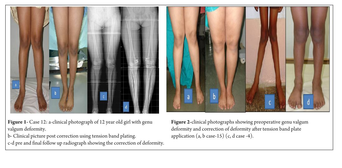
Figure 1, 2

Surgical technique
With patient in supine position, tourniquet was applied to achieve haemostasis. Centre of Physis was marked using k wire under fluoroscopy guidance. Incision is taken and dissection done to reach the periosteum taking care that the periosteum is not breached and perichondrial blood supply is maintained. Plate inserted over the K wire and position confirmed under image intensifier, cannulated screws inserted parallel to Physis. Screw position checked under image intensifier in both antero-posterior and lateral view. Closure done and dressing applied. Postoperatively no immobilisation was required and patients were mobilised full weight bearing as tolerated from post op day 1. No walking support was required. Patients were followed up prospectively at every 3 month and knee radiographs were taken antero-posterior and lateral view to look for screw divergence and clinically IMD/ICD was measured.

Figure 3

Results
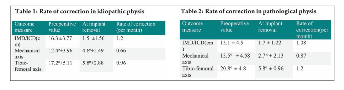
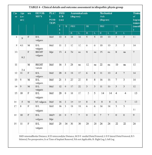
The average age of intervention was 7.4±2.96 years (range 2.4 -11.2years).The mean values of mechanical axis, tibiofemoral axis, IMD/ICD and duration of correction were calculated excluding the patient with overcorrection [case no 19] to avoid wide deviation from mean values. The mean preoperative IMD/ICD was 15.8cm ± 3.96 (range 10 cm to 22 cm) .The mean correction 1.6cm ± (range 0cm to 4 cm). Rate of correction was 1.14 cm per month. The mean preoperative mechanical axis was 13.4o± 5 (range7o to 25o).The mean post operative mechanical axis was 3.9o ±2.44 (range 0o to 10o). Rate of correction was 0.76 o per month .The mean preoperative tibiofemoral angle was 18.7o ±5.1 (range 7o -28o). The mean post operative tibiofemoral angle was 5.8o ± 2.29 (range 0o -12o). Rate of correction was 1.04o per month .Rate of correction in idiopathic and pathological physis group are depicted in table 1 and 2).The average duration of eight plate removal was 12.4 months (range 7-24 months). It was 13.3 months in pathological group and 11.8 months in idiopathic group. Figure 1 shows clinical and radiological correction in terms of IMD/ICD, mechanical axis and tibiofemoral axis. One patient was excluded from the data, a case of pathological genu valgum because the physis became fused before correction was achieved .The patient was treated with corrective osteotomy. Thus, emphasizing on preoperative planning in terms of age of intervention and aetiology deformity. There were two complications; one patient had screw back out which was revised. Other patient had reversal of deformity from 24° valgus to 22° varus due to delayed follow up [case 19]. She was treated with removal of 8 plate and is currently under observation for spontaneous correction. None of the patient had limb length discrepancy except (case 12) had 3 cm femoral shortening. Clinical data and outcome assessment in idiopathic and pathological group are depicted in table 1 and table 2.

Table 1, 2

Discussion
Physeal growth in child depends on variety of factors like biomechanical, hormonal and genetic [17]. Growth modulation using eight-plate depends on biomechanical growth modification based on Hueter-volkmann principle. Sustained compression parallel to physis leads to growth retardation and subsequent correction of deformity [9]. Eight-plate functions as flexible device which produces sustained compression at physis. The compression is not constant as the screws diverge with correction and with maximum divergence the plate bends, hence also called as tension band plate [11]. Eight-plate serves as non rigid implant with lateralisation of fulcrum for deformity correction. Thus, leading to faster rates of correction [ 2, 8]. Staples and transphyseal screws are rigid implants with centralised fulcrum for deformity correction [13]. They produce constant compression at physis. Thus they take longer time for deformity correction [2, 8]. Staples and transphyseal screws are rigid implants and if a prolonged duration is required for correction of deformity, they may cause physeal arrest [17]. In contrast eight plates are relatively flexible implants as it allows for screw separation. This is one of the reasons for decreased incidence of physeal arrest and makes it safer to use in younger children when compared to staples. In our study, three patients below the age of 3 years were treated successfully without any complications [case no 2, 7 and 11]. Clinical assessment of growth modulation by using IMD/ICD is reported for studies using staples [18] but not in studies using eight-plate. Since IMD/ICD was the major criteria to define the indication for surgery in our series, we have used the same as the primary outcome measure. We believe that IMD/ICD is an important clinical measure as majority of our patient were asymptomatic with cosmetic deformity and follow up parent counselling was easier. However standard values for particular age and race vary [14, 19].There can also be high rate of inter observer discrepancy and this is one of the limitations of the study. Radiographic measurements are used as outcome measures in papers on growth modulation using eight-plate like tibio-femoral axis, mechanical axis, joint orientation angles, mechanical axis deviation , articular –diaphyseal etc[2,10,11,20]We have used two radiological outcome measures, the hip knee ankle mechanical axis and the tibio-femoral angle. In our study we found that tibiofemoral angle and mechanical axis improved significantly. Standard antero-posterior and lateral radiographs of knee joint are useful in follow ups to see the effect of epiphysiodesis as seen with screw divergence. Joint orientation angles were not used, as scanograms become distorted due to magnification and parallax leading to false values[6].Also in young children it is difficult to visualise the distal femur and proximal tibia epiphyseal contours to accurately mark these angles. Ballal et al [11] showed that mean rate of correction of tibiofemoral axis 0.7°/month for distal femur and 0.5°/month in proximal tibia. Burghardt et al [20] showed mechanical axis correction of 1.73mm/month. In our study mean rate of correction of mechanical axis was 0.76°/month and tibio-femoral axis was 1.04°/month.

Table 3

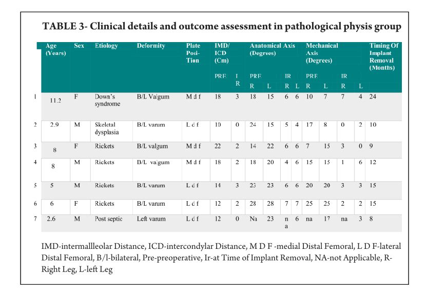
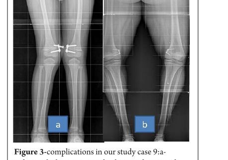
Rate of correction is faster in children less than 10 years as shown in study by Ballal et al [11]. In our study, due to smaller sample size of patients more than 10 years of age we could not assess this in our series. We did compared the rate of correction between the idiopathic and pathological group with former showing faster correction (Table 3 and 4). This corroborated with findings of Boero et al.No difference in rate of correction was encountered in terms of gender and type of deformity in our series. In our study two complications occurred, one patient had a screw back out (figure 3a) at 4 month after insertion, which was treated with revision of screw. However the rate of deformity correction was consistent with other patients in idiopathic physis group. We believe that the reason for back out may be placement of screw near the posterior cortex. Similar complications of screw loosening were seen in studies by Burghardt (n=1) [20] and Stevens (n=1) [2]. Other complication of overcorrection of deformity (figure 3b) was seen in one patient because of delayed follow up. This is justified as principally the sustained compression at physis will produce dynamic changes. Ballal et al [11] encountered one case of both screw and plate migration which was revised, such complication didn’t occur in our series. Several series encounter rebound growth after plate removal [2, 11, 20], we have not seen rebound of deformity in our patient with longest follow up of 2 years after plate removal. Complications related to implant like migration, breakage, bending of implants etc seen with staples are less common with eight -plate. Most dreaded complication of physeal arrest is not reported in our and other series [2, 3, 7, 10, 11, 20]. The threaded screws are less likely to extrude, especially in cartilaginous physis seen in younger age [8]. Screws are placed extra-periosteal and perichondrial blood supply is not hampered, so the chances of physeal arrest while insertion and removal are very less. Our study has drawbacks of small sample size and retrospective study design. However it does gives important inferences in terms of ability of 8 plates to correct the deformity and more importantly with minimal complications. However larger sample comparative studies will be required to establish the superiority of this method compared to other methods.  Each author certifies that he or she has no commercial associations (eg, consultancies, stock ownership, equity interest, patent/licensing arrangements, etc) that might pose a conflict of interest in connection with the submitted article.

Table 4


References

1. Celestre PC, Bowen RE. Correction of angular deformities in children- Current Orthopaedic Practice. 2009;20(6):641-647.
2. Stevens PM. Guided growth for angular correction: a preliminary series using a tension band plate. J Pediatr Orthop. 2007 Apr-May; 27(3):253-9.
3. Wiemann JM 4th, Tryon C, Szalay EA. Physeal stapling versus 8-plate hemiepiphysiodesis for guided correction of angular deformity about the knee. J Pediatr Orthop. 2009 Jul-Aug; 29(5):481-5.
4. Cho TJ, Choi IH, Chung CY, Yoo WJ, Park MS, Lee DY. Hemiepiphyseal stapling for angular deformity correction around the knee joint in children with multiple epiphyseal dysplasia. J Pediatr Orthop. 2009 Jan-Feb; 29(1):52-6.
5. Ghanem I, Karam JA, Widmann RF. Surgical epiphysiodesis indications and techniques: update. Curr Opin Pediatr. 2011 Feb; 23(1):53-9.
6. Friend L, Widmann RF. Advances in management of limb length discrepancy and lower limb deformity. Curr Opin Pediatr. 2008 Feb; 20(1):46-51.
7. Burghardt RD, Herzenberg JE, Standard SC, Paley D. Temporary hemiepiphyseal arrest using a screw and plate device to treat knee and ankle deformities in children: a preliminary report. J Child Orthop. 2008 Jun; 2(3):187-97.
8. Eastwood DM, Sanghrajka AP. Guided growth: recent advances in a deep-rooted concept. J Bone Joint Surg Br. 2011 Jan; 93(1):12-8.
9. Stevens PM. Guided growth of the lower extremities. Current Orthopaedic Practice. March/April 2011; 22(2):142–149
10. Schroerlucke S, Bertrand S, Clapp J, Bundy J, Gregg FO. Failure of Orthofix eight-Plate for the treatment of Blount disease. J Pediatr Orthop. 2009 Jan-Feb; 29(1):57-60.
11. Ballal MS, Bruce CE, Nayagam S. Correcting genu varum and genu valgum in children by guided growth: temporary hemiepiphysiodesis using tension band plates. J Bone Joint Surg Br. 2010 Feb; 92(2):273-6.
12. Stevens PM, Klatt JB. Guided growth for pathological physes: radiographic improvement during realignment. J Pediatr Orthop. 2008 Sep; 28(6):632-9.
13. DeBrauwer V, Moens P. Temporary hemiepiphysiodesis for idiopathic genuavalga in adolescents: percutaneous transphyseal screws (PETS) versus stapling. J Pediatr Orthop. 2008 Jul-Aug; 28(5):549-54.
14. Heath CH, Staheli LT. Normal limits of knee angle in white children–genu varum and genu valgum. J Pediatr Orthop. Mar-Apr 1993.
15.Paterson HA Epiphyseal growth plate fracture. Springer, Berlin 2007.
16. Paley D. Principles of Deformity Correction. Berlin, Germany: Springer; 2002.
17. Frost HM, Schönau E. On longitudinal bone growth, short stature, and related matters: insights about cartilage physiology from the Utah paradigm. J Pediatr Endocrinol Metab. 2001 May; 14(5):481-96.
18. Courvoisier A, Eid A, Merloz P. Epiphyseal stapling of the proximal tibia for idiopathic genu valgum. J Child Orthop. 2009 Jun; 3(3):217-21.
19. Omololu B, Tella A, Ogunlade SO, Adeyemo AA, Adebisi A, Alonge TO, Salawu SA, Akinpelu AO. Normal values of knee angle, intercondylar and intermalleolar distances in Nigerian children. West Afr J Med. 2003 Dec; 22(4):301-4.
20. Burghardt RD, Herzenberg JE. Temporary hemiepiphysiodesis with the eight-Plate for angular deformities: mid-term results. J Orthop Sci. 2010 Sep; 15(5):699-704.
21. Boero S, Michelis MB, Riganti S. Use of the eight-Plate for angular correction of knee deformities due to idiopathic and pathologic physis: initiating treatment according to etiology-J Child Orthop 2011;5(3):209-216.

How to Cite this Article: Patwardhan S, Shah K, Shyam AK, Sancheti P. Growth Modulation in Children for Angular Deformity Correction around knee – Use of Eight Plate. International Journal of Paediatric Orthopaedics July-Sep 2015;1(1):33-37.        

(Abstract)      (Full Text HTML)      (Download PDF)


Nerve Injuries and Myositis Ossificans associated with Supracondylar Humerus Fracture

Vol 1 | Issue 1 | July-Sep 2015 | page:30-32 | Maulin Shah, Maulik Patel.


Authors : Maulin Shah[1], Maulik Patel[2].

[1] Consultant Pediatric Orthopedic Surgeon, Orthokids Clinic, Ahmedabad, India.
[2] Clinical Fellow , Orthokids Clinic, Ahmedabad, India.

Address of Correspondence
Dr. Maulin Shah
OrthoKids Clinic,
Kamdhenu House, Opp. Apang Manav Mandal,
Drive-in Road, Memnagar, Ahmedabad – 380 052.
Email : orthokidsclinic@gmail.com


Abstract

Background: Angular deformities around the knee joint in skeletally immature children are treated with methods of reversible hemiepiphysiodesis like staples, transphyseal screw and eight plate. Hemiepiphysiodesis using Eight plate has showed good results with advantage being faster correction, less complications and can be used in younger age.
Methods: The aim of this retrospective study is show the efficacy of eight plate application and its complication rate. Nineteen patients (37 physes) (unilateral: 3; bilateral: 16) with angular deformity were treated with eightplate application. Seven with pathological physes and twelve with idiopathic physes. Outcome assessment was done clinically with calculation of intermalleolar /intercondylar distance and radiologicaaly with mechanical and anatomical axis. Correction achieved was considered when anatomical/mechanical axis were within normal limits and intermalleolar/intercondylar distance was less than 5 cm.
Results: The average age of intervention was 7.4±2.96 years (range 2.4 -11.2years). Rate of correction of IMD/ICD was 1.14 cm per month. Rate of correction of mechanical axis was 0.76 o per month. Rate of correction of anatomical axis was 1.04o per month. The average duration of eight plate removal 12.4 months (range 7-24 months).There were two complications one patient with screw backout and other with overcorrection.
Conclusion: Reversible hemiepiphysiodesis using eight plate is and effective method with minimal complications and faster rates of correction. Idiopathic physes show faster rates of correction than pathological physes. Physeal growth arrest is not seen with eight plate application. Larger data and long term follow up is required to assess the rebound deformity after eight plate removal.
Keywords: Reversible, hemiepiphysiodesis, angular deformity, eight plate.


Introduction
Supracondylar humerus fractures are the most common upper limb injury amongst children. Due to the vicinity of the important nerves in this area, nerve injuries are frequently encountered with these fractures.

Incidence
Incidence of nerve injuries associated with supracondylar humerus fractures is reported to be 10 to 20% in different published studies. Most of these injuries can be identified preoperatively by proper clinical evaluation. Anterior interosseous nerve is the most commonly encountered nerve injury in extension type of fractures with incidence of 12 to 15%.[1-3] Radial nerve injury is second most common in this group with occurrence about 8%. Ulnar nerve injury is least commonly seen in extension injuries & approximate incidence is about 3%[4,5]. Flexion type of supracondylar fractures have more common association with Ulnar nerve injury.
Fracture morphology & Nerve Injuries
In extension type of fractures, median nerve is injured with postero-lateral displacement & radial nerve is injured with postero-medial displacement of fracture. In a study of 59 consecutive cases of type -III fractures, Crowford and collegues reported that 87% of median nerve injuries were associated with postero-lateral displacement and all radial nerve injuries were associated with postero-medial displacement[6]. The rate of acute neurologic injury in ipsilateral supracondylar humerus and forearm fractures is almost twice than that found in patients with isolated supracondylar humerus fractures. In a series of 150 patients with ipsilateral injuries, Muchow et. al. observed that the overall incidence of nerve palsy was 18.9% when a forearm fracture required reduction compared with only 7.3% in a forearm fracture that was not reduced[7]. In a series of 26 open supracondylar humerus fractures, Ozkul et. al. reported the incidence of nerve injury to be as high as 34%. A careful pre-operative evaluation to identify the nerve injury is thus advocated in open injuries[8]. Correlation was also found between severity of fracture type and incidence of nerve injury. Gartland type- II had 7%, type -III had 19% and type-IV had 36% chances of nerve injuries[9]. Nerve interposition between fracture fragments can cause failure of closed reduction. In a series of 41 failed closed reduction, Fleuriau-Chateau et. al. reported that 15 patients had entrapment of Median Nerve or Radial Nerve. Thus, incidence of nerve injury in failed closed reduction is approximately 35%[10].

Clinical Evaluation for Nerve Injury
Although it is difficult to do complete neurological evaluation in an injured child, it is very important to note the status of pre-operative movements. Trainees should note the ability of a child to carry out specific tasks rather than pointing specific nerve injuries. Median nerve injury can be identified by loss of thumb & index finger inter-phalangeal flexion. “Pointing index finger” while the child is asked to flex the fingers is a cardinal sign. Loss of extension at Metacarpo-phalangeal joints of fingers & thumb extension is suggestive of Radial Nerve involvement. Clawing of ulnar fingers & loss of adduction-abduction of fingers suggests Ulnar Nerve injury. Sensory deficits are difficult to indentify in acute injuries[11].

Types of Nerve Injuries
Most of the nerve injuries associated with supracondylar fractures are neuropraxic in nature. It is produced by the perineural fibrosis induced by direct compression of the fractured bony fragment. These injuries have good potential of spontaneous recovery. Complete transaction of the nerve is rare and radial nerve is the most commonly involved in this category[12]. Nerve can get entrapped in callus and produce symptoms of nerve injury. Radiologically, it gives appearance of a hole in the bone & is known as “Metev’s Sign”[3]. Compartment syndrome is an uncommon but known complication of Supracondylar fracture in today’s era. Increased pressure in the compartment causes nerve ischemia. Median Nerve paresis is the most commonly observed in this category. Majority of Ulnar Nerve injuries are caused Iatrogenically rather than direct injury.

Iatrogenic Nerve Injuries
Ulnar nerve injury is observed more as iatrogenic injury rather than post traumatic injury. Recent literature reports iatrogenic Ulnar nerve injury incidence to be 3% – 4%[13]. Cross pinning configuration has shown more chance to contracting Ulnar nerve injury compared to lateral pinning.[14] Passage of Ulnar nerve between Olecranon and the medial epicondyle makes it more prone to injure when medial pin I s passed in the cubital groove. Skaggs et. al. reported that hyperflexion of elbow increases chance of Ulnar nerve injury threefold & they recommend reducing elbow flexion after placing the first lateral pin in extension variety of supracondylar fractures[9]. Majority of instances the Ulnar nerve is injured by direct trauma or compression of sheath due to winding while drilling of wire. Shtarker et. al. used Ulnar nerve monitoring through electrical stimulation during & at the end of medial pinning. They found this technique safe, simple & easily applicable. In a series of 138 patients, by using this method, they did not notice any Ulnar nerve injury after doing crossed pinning[15]. Mulpuri et. al. found a mini-open technique useful to reduce the incidence of iatrogenic Ulnar nerve injuries while doing cross pinning. They recommended retraction of Ulnar nerve under direct vision by a 1.5 cm medial incision before introducing medial pin[16]. Literature reports that about 17-30% children have Ulnar nerve instability. An ultrasound study of the Ulnar nerve anatomy done by Eraze & colleagues, suggested that the incidence of anterior subluxation & dislocation of the Ulnar nerve is significantly high in patients with generalized hyperlaxity compared to normal population. They suggested that the ultrasound evaluation and assessment of ligamentous laxity are additional tools which can identify children at risk of iatrogenic nerve injury[17]. Iatrogenic radial nerve injury is rare and associated with piercing by medial pin as it exists through the anterolateral cortex. Most of these injuries are neuropraxia and spontaneous recovery occurs usually. Medial pin penetration in the opposite cortex should be limited to 1 mm to 2mm to prevent radial nerve injury[18].

Treatment approach to Nerve Injuries
Most nerve injuries are neuropraxias in nature & they generally show spontaneous recovery in 3 months time. Franklin et. al. suggested need of immediate exploration in nerve palsy with accompanying pulselessness[12]. Reducible fractures with nerve injuries should be treated with closed reduction and close follow up. Irreducible fractures with nerve deficits require open reduction to rule out nerve entrapment. If nerve deficit is found within a few hours of cross pinning then pin should be removed and nerve should be explored. It is advisable to change to lateral pinning. Nerve deficit after a few weeks of cross pinning can be managed by pin removal and observation for 5 to 6 months. If recovery does not occur then neurolysis is the mainstay of treatment. Rarely nerve grafting is indicated.

Conclusion
Nerve injuries associated with supracondylar humerus fracture is a frequent occurrence. One should have high degree of suspicion about it & a careful pre-operative clinical examination is needed to report it. Median nerve injury is the most common nerve injury associated with extension variety of fractures. Incidence significantly increases in open fractures, ipsilateral forearm fractures & fractures with vascular compromise. Ulnar nerve injury is commonly happen as iatrogenic injury due to cross pinning. A mini-open technique or ultrasound evaluation or electric nerve monitoring during surgery are recommended tools to reduced its incidence. Most of the nerve injuries are neuropraxias and they spontaneously resolve by 6 months. Small percentage of patients may need nerve exploration & repair.

Myositis ossificans in Supracondylar humerus fractures
Wilkins reported 1.4% incidence of myositis ossificans in his meta-analysis of 470 cases of supracondylar humerus fractures[19]. Brachialis is the commonest muscle to get involved and causes restriction of range of motion[20]. High energy trauma, manipulation by bone setter, aggressive postoperative physiotherapy and overzealous dissection while open reduction are the risk factors[21]. In early stage, patients present with pain, redness, local warmth and swelling. In late stage, once the ossification settles, bony mass is palpable separately from the underlying bone and it causes restriction of motion at the elbow[22]. On plain radiographs, myositis looks like calcification in its early stages. Mature myositis mass demonstrates well defined outer shell of the bone, commonly at anterolateral aspect of the elbow. CT scan is helpful to confirm[23]. Early stage is treated by analgesics & anti-inflammatory medicines and restriction of passive exercises. Mature myositis mass should be excised completely. Material should be sent for the histopathological examination[22,24]. Preoperative or early postoperative radiotherapy has been reported to prevent myositis ossificans occurrence in at-risk patients. Prophylactic dose should be between 600 and 1000 cGy[25, 26].


References

1. Cramer KE, Green NE, Devito DP. Incidence of anterior interosseous nerve palsy in supracondylar humerus fractures in children. Journal of Pediatric Orthopaedics. 1993;13(4):502-505.
2. Spinner M, Schreiber SN. Anterior interosseous-nerve paralysis as a complication of supracondylar fractures of the humerus in children. Journal of Bone Joint Surgery (Am). 1969; 51(8):1584-1590.
3. McGraw JJ, Akbarnia BA, Hanel DP, et al. Neurological complications resulting from supracondylar fractures of the humerus in children. Journal of Pediatric Orthopaedics.1986; 6(6):647-650.
4. Skaggs DL, Hale JM, Bassett J, et al. Operative treatment of supracondylar fractures of the humerus in children. The consequences of pin placement. Journal of Bone Joint Surgery (Am). 2001; 83A(5):735-740.
5. Cheng JC, Lam TP, Shen WY. Closed reduction and percutaneous pinning for type III displaced supracondylar fractures of the humerus in children. Journal of Orthopaedic Trauma. 1995; 9(6):511-515.
6. Crowford CC, Peters WM, John EB, James KR, Michael MB. Neurovascular injury and Displacement in Type III Supracondylar Humerus Fractures. Journal of Pediatric Orhtopaedics. 1995 Jan:Feb ;15(1) : 47-52
7. Muchow RD, Riccio AI, Garg S, Ho CA, Wimberly RL. Neurological and vascular injury associated with supracondylar humerus fractures and ipsilateral forearm fractures in children. Journal of Pediatric Orthopaedics. 2015 March;35(2):121-5.
8.Ozkul E , Gem M, Arslan H, Alendar C, Demirtas A, Kisin B. Surgical treatment outcome for open supracondylar humerus fractures in children. Acta Orthopaedics Belgium. 2013 October;79(5):509-513.
9. Joiner ER, Skaggs DL, Arkader A, Andras LM, Lightdale-Miric NR, Pace JL, Ryan DD. Iatrogenic nerve injuries in the treatment of supracondylar humerus fractures: are we really just missing nerve injuries on preoperative examination? Journal of Pediatric Orthopaedics. 2014 June; 34(4):388-92.
10. Fleuriau-Chateau P, McIntyre W, Letts M. An analysis of open reduction of irreducible supracondylar fractures of the humerus in children. Canadian Journal of Surgery. 1998; 41(2):112-128.
11. RW Culp, AL Osterman, RS Davidson, T Skirven, FW Bora. Nerural injuries associated with supracondylar fractures of humerus in children. Journal of Bone Joint Surgery.1990 Sep; 72(8):1211-1215.
12. Franklin CC, Skaggs DL. Approach to the pediatric supracondylar humeral fracture with neurovascular compromise. Instructional Course Lectures. 2013;62:429-33.
13. Cheng JC, Ng BK, Ying SY, Lam PK. A 10-year study of the changes in the pat- tern and treatment of 6,493 fractures. Journal of Pediatric Orthopaedics. 1999;19:344-50.
14. Wilkins KE. Fractures and dislocations of the elbow region. In: Rockwood CA Jr, Wilkins KE, King RE, editors. Fractures in children. Volume 3. 3rd edition. New York: JB Lippincott; 1991. p 526-617.
15. Shtarker H, Elboim-Gabyzon, Bathish E, Laufer Y, Rahamimov N, Volpin G. Ulnar nerve monitoring during percutaneous pinning of supracondylar fractures in children. Journal of Pediatric Orhtopaedics. 2014March;34(2):161-5.
16. Mulpuri K, Tritt BL. Low incidence of ulnar nerve injury with crossed pin placement for pediatric supracondylar humerus fractures using a mini-open technique. Journal of Orthopaedic Trauma. 2006 March;20(3);234
17. Erez O, Khalil JG, Legakis JE, Tweedie J, Kaminski E, Reynolds RA. Ultrasound evaluation of ulnar nerve anatomy in the pediatric population. Journal of Pediatric Orthopaedics. 2012 September ;32(6):641-6.
18. Sairyo Koichi, Henmi Tatsuhiko, Kanematsu Yoshiji, Nakano Shunji, Kajikawa Tomomasa. Radial Nerve Palsy Associated with Slightly Angulated Pediatric Supracondylar Humerus Fracture. Journal of Orthopaedic Trauma. 1997 April;11(3):227-229.
19. Wilkins KE. Fractures and dislocations of the elbow region. In: Rockwood CA, Wilkins KE, King RE. Fractures in Children. 3rd edition. New York: JB: Lippincott; 1991; 509-28.
20. Naranje S1, Kancherla R, Kannan A, Malhotra R, Sharma L, Sankineani SR. Extraarticular bony ankylosis in a child with supracondylar fracture of humerus. Chinese Journal Traumatology. 2012;15(5):300-2.
21. Hartigan BJ1, Benson LS. Myositis ossificans after a supracondylar fracture of the humerus in a child. American Journal of Orthopedics. 2001 Feb;30(2):152-4.
22. Spinner, Robert J.; Jacobson, Scott R.; Nunley, James A. Case Report Fracture of a Supracondylar Humeral Myositis Ossificans. Journal of Orthopaedic Trauma: June 1995; 183-277
23. Zeanah WR, Hudson TM. Myositis ossificans: radiologic evaluation of two cases with diagnostic computed tomograms. Clinical Orthopedic Related Research. 1982 Aug;(168):187-91.
24. Augustus Thorndike Jr. Myositis ossificans traumatica. Jounal of Bone Joint Surg Am, 1940 Apr; 22 (2): 315 -323.
25. Brady LW. Radiation-induced sarcomas of bone. Skeletal Radiology 1979;4:72-8.
26. Kim JH, Chu FC, Melamed HQ, Huvos A, Cantin J. Radiationinduced soft-tissue and bone sarcoma. Radiology 1978;129:501-8.


How to Cite this Article: Shah M, Patel M. Nerve Injuries and Myositis Ossificans associated with Supracondylar Humerus Fracture. International Journal of Paediatric Orthopaedics July-Sep 2015;1(1):30-32.         

 Photo


(Abstract)      (Full Text HTML)      (Download PDF)