Tag Archive for: management

Supracondylar Humerus Fractures in Children: Epidemiology and Changing Trends of Presentation

Vol 1 | Issue 1 | July-Sep 2015 | page:3-5 | Sandeep V Vaidya.


Authors : Sandeep V Vaidya[1,2,3*].

[1] Children’s Orthopaedic Clinic, Thane.
[2] B J Wadia Children’s Hospital, Parel Mumbai
[3] Jupiter Hospital, Thane, Maharashtra, India

Address of Correspondence
Dr Sandeep V Vaidya
Director, Children’s Orthopaedic Clinic, Thane. India.
Email: drsvvaidya@gmail.com


Abstract

Diseases show a tendency to vary according to changing socio-economic trends and fractures too have shown this tendency. Paediatric supracondylar humerus fractures are one of the most common fractures seen by paediatric orthopaedic surgeons. There are few notable trends that have been reported and few other that I have personally noted in my practice and in practice of my colleagues. This article put together the changes reported in literature and tries to combine it with clinically relevant practical situations. Special focus is on fracture presentation and on decision making in management.
Keywords: Supracondylar Humerus fracture, classification, management.


Introduction:
Supracondylar humerus fractures in children are commonly seen in day to day practice. In this section, we study the epidemiology and changing trends of these fractures with respect to incidence, patient profile, types, modes of injury, treatment trends and complications.

Incidence:
Supracondylar humerus fractures (SHF) comprise 17% of all pediatric fractures and are second in frequency to forearm fractures. According to an epidemiological study, the incidence of fracture supracondylar humerus is 308/100000 per year in the general population. It is also the commonest pediatric fracture around the elbow. One epidemiological study identified supracondylar fractures in 206 out of 355 elbow fractures (58%) [1]. Barr reported a higher incidence of supracondylar humerus fractures during the vacations [2].

Age and sex:
If age distribution is considered, in the 0 to 7 year age group, SHF is easily the commonest fracture seen (28%) [3]. The mean age at which fracture supracondylar humerus occurs is 5 to 8 years [1,2]. Wilkins proposed that when a child falls on extended upper extremity, the patients who demonstrate hyperextension (cubitus recurvatum) of the elbow are more predisposed to have supracondylar fractures. The children who do not have hyperextension of the elbow tend to sustain fractures of the radius and the ulna, usually at the distal portion. Since ligamentous laxity with elbow recurvatum is seen in younger children, this explains the higher incidence of supracondylar humerus fractures in younger children and higher incidence of radius ulna fractures in older children.
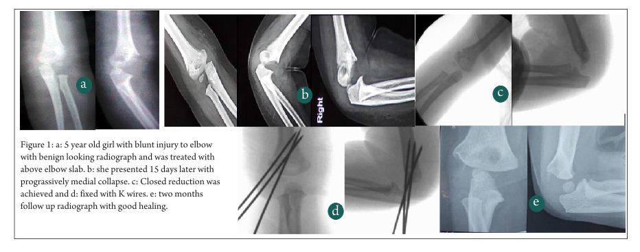
Recently, there seems be increase in incidence of SHF in lower age group(less than 2 years). Fractures occurring in these very young children may pose a diagnostic dilemma because in many of these cases, the fracture line is extremely low and on plain radiographs may mimic a fracture lateral condyle humerus due to the largely cartilaginous component of the distal fragment. In such cases, additional imaging like MRI or arthrogram may be needed to differentiate these low supracondylar fractures from the lateral condyle fractures (Fig. 1). Another peculiarity of the low supracondylar humerus fractures is that such fractures can be complicated by Avascular necrosis of the trochlea with subsequent later sequelae.

Fig 1

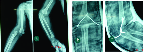
Figure 1: (Case Courtesy Dr Sandeep Patwardhan)1a: Elbow radiograph`of a 2 year old child with fall on outstretched hand. The fracture line is extremely distal and only a flake of metaphysis is seen.1b,c: The fracture was treated by closed reduction and K wire pinning .

In most of the earlier studies, the fracture occurred much more commonly in boys than in girls. However in most of the recent series, the frequencies in girls and boys seems to be equalizing. Some series have actually reported a higher incidence in girls than boys[1,2]. This changing sex distribution may be attributed to more active participation of girls in sports activities.

Mode of injury:
The cause of fracture supracondylar humerus is accidental fall while playing in most of the cases (60 to 80 %). Road traffic accidents account for 10 to 20% of SHF [2]. High velocity trauma can lead to fractures with metaphyseal comminution or in rare cases fractures with intercondylar extension.
Child abuse is an uncommon etiology of SHF[4]. However Strait and colleagues reported supracondylar fractures from abuse in three of 10 abused children under the age of 3, and cautioned that SHF should not be assumed to have non-abusive causes without careful consideration [5].

Types:
Extension type is the commonest type, flexion type is seen in 1 to 3% cases [6]. The patients in the flexion-type group (mean age, 7.5 years) are significantly older than those in the extension-type group (mean age, 5.8 years). The fractures in flexion-type group are also more probable to require open reduction (31%) than those in the extension-type group (10%). The flexion-type group had a significantly increased incidence rate of ulnar nerve symptoms (19% vs 3% in the extension-type group) and need for ulnar nerve decompression [7].
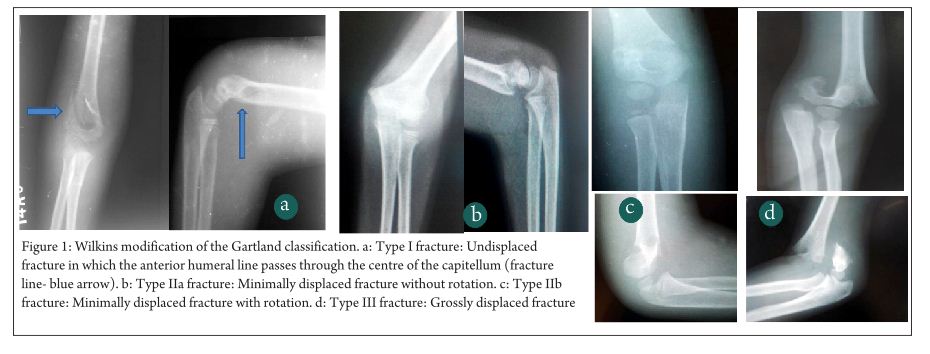
Gartland classification is the commonest classification system used to grade supracondylar humerus fracture. Grade 1 fractures are the commonest, followed by Grade 2 and then Grade 3 [1,2].
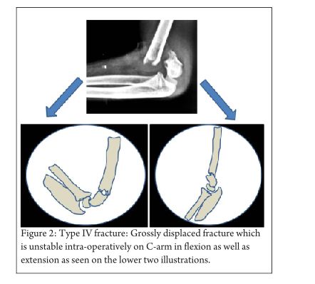
In addition to these 3 types, Leitch et al described a type 4 fracture with multidirectional instability (unstable in both flexion and extension). This fracture type was noted in 9 out of 297 displaced fractures. These fractures are associated with high velocity trauma, the periosteal sleeve is completely torn and special manoeuvres are needed for closed reduction- pinning [8].
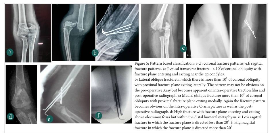
In extension type fractures the distal fragment may be displaced posteromedially or posterolaterally. Posteromedial displacement is commoner and seen in approximately 75% cases in most series. Posteromedial displacement of the distal fragment places the radial nerve at risk, whereas in fractures with posterolateral displacement the brachial artery and median nerve are at risk [9]. Bahk et al additionally classified extension type supracondylar fractures based on orientation of the fracture line in coronal as well as sagittal planes. In coronal plane, transverse fractures were the commonest (49%) followed by lateral oblique fractures (44%). Medial oblique (4%) and high transverse fractures (3%) were less common. Whereas transverse and lateral oblique fractures are amenable to lateral only pinning, the medial oblique and transverse fractures need to be fixed with medial-lateral cross pins [10].
High SHF are also being increasingly reported recently. Sen et al reported an incidence of high metaphyseal- diaphyseal supracondylar humerus fractures in 6 out of 182 fractures [11].

Treatment:
Blount in 1955 had cautioned against operative treatment in SHF citing the high incidence of complications following operative treatment [12]. However with significant advances in operative techniques and intraoperative imaging, operative treatment with Closed Reduction Percutaneous Pinning (CRPP) is easily the treatment of choice for displaced supracondylar humerus fractures [13]. Approximately 40% of SHF are treated operatively making it the commonest pediatric fracture to undergo operative treatment [2]. Cheng et al in an epidemiological study of 6493 fractures reported that the closed-reduction and percutaneous pinning rates for supracondylar humerus fractures increased 4.3 to 40% over a 10 year period from 1985 to 1995. The changes in treatment pattern were also accompanied by a corresponding decrease in the open-reduction rate and hospital stay periods from <10% to 38% of patients being discharged within 1 day of admission in the 10-year period [3].

The incidence of operative treatment is 0% in Grade 1 fractures, almost 50% for Grade 2 fractures, 100% for Grade 3 fractures and 100% for flexion type fractures. The incidence of open reduction is highest in flexion type fractures (50%) [2]. In an epidemiological study, out of 3235 children with displaced SHF treated operatively at a tertiary care children’s hospital at Toronto, 78.7% underwent operative treatment in the form of Closed Reduction Percutaneous Pinning (CRPP) whereas the remainder 21.8% underwent Open Reduction Internal Fixation (OR). There was a significant difference in the delay to surgery between the CRPP and OR groups [14]. In developed countries, there is a trend for more number of SHF are being treated by pediatric orthopaedic subspecialists. In New England, only 37% of SHF were treated by Pediatric Orthopaedic surgeons in 1991, this number rose to 68% in 1999. Kasser et al reported that in fractures treated by pediatric orthopaedic surgeons the length of hospitalization was lesser (1.4 ± 0.4 days) than for fractures treated by general orthopaedic surgeons (2.2 ± 0.6 days) [15]

Pin configurations, changing trends:
Pin configurations used by surgeons have shown a changing trend over the past decade. Several biomechanical studies published before 2005 revealed that crossed medial- lateral pin configurations are biomechanically stronger than lateral only pin configurations. Hence crossed medial- lateral pinning was preferred. However a major danger of the medial pin was iatrogenic ulnar nerve injury. Incidence of iatrogenic ulnar nerve injury with crossed medial- lateral pinning in various series has ranged from 0% to 6% [16,17]. Lyons et al reported iatrogenic ulnar nerve palsy in 19 out of 375 crossed medial- lateral pinning. 15 out of these 19 palsies recovered within 4 months after medial pin removal. However 4 palsies failed to recover, underwent ulnar nerve exploration and neurolysis [17]. A systematic pooled analysis of 32 trials comprising 2639 children suggests that there is an iatrogenic ulnar nerve injury for every 28 patients treated with the crossed pinning compared with the lateral pinning [16].
An inherent fallacy of the early biomechanical studies was that these studies were based on in-vitro findings wherein loads applied to create displacement were significantly higher than those which would be applied in-vivo wherein the fixation would be additionally supplemented with plaster slab application. Lee et al in their series of 61 consecutive lateral only pinning reported a zero incidence of loss of reduction as well as iatrogenic ulnar nerve palsy [18]. A randomized controlled study published in 2007 concluded that lateral entry only pinning did not result in increase incidence of loss of reduction as compared to crossed medial-lateral pinning [19]. A survey involving eight surgeons conducted in 2012 confirmed that this RCT had a significant influence on the surgeons’ practice. Five out of eight surgeons individually had a statistically significant change in their practice pattern for pin configuration. Except for certain selected fracture patterns, lateral only pinning is being increasingly used as the standard pin configuration for supracondylar humerus fractures [20].

Complications:
Complications of fracture supracondylar humerus include compartment syndrome, vascular injury, nerve injury (fracture related or iatrogenic) and malunion with cubitus varus deformity. The incidence of compartment syndrome is approximately 0.1% to 0.3% of all supracondylar humerus fractures [21]. Ipsilateral forearm fracture significantly increases risk of compartment syndrome [22]. In a study, the incidence of compartment syndrome was — % in fractures reduced and fixed within – hours of injury as compared to — % in fractures fixed after a delay of – hours.
The incidence of vascular injuries is approximately 20% and majority are associated with Grade 3 fractures [1, 23, 2]. Fractures with posterolateral displacement are more at risk for vascular injuries (approximately 65%) than fractures with posteromedial displacement (approximately 53%) [23]. If the hand is well perfused but pulseless, the great majority of the time fracture reduction is sufficient treatment. In contrast, patients presenting with a pulseless and poorly perfused hand have a nearly 50% chance of requiring vascular surgery and nearly 25% chance of developing a compartment syndrome [24, 25].
Nerve injuries are seen in approximately 4% fractures and majority are associated with Grade 3 fractures [1,2]. Overall, the most commonly injured nerve is median nerve (50%) followed by radial nerve (28%) followed by ulnar nerve (22%). The pattern of displacement is the most important risk factor in nerve injury. In fractures with median nerve palsy, posterolateral displacement is seen in 87% cases. In cases with radial nerve palsy, posteromdeial displacement Is seen in almost all cases [23] In flexion type, ulnar nerve is most commonly injured [7].


References

1. Houshian S, Mehdi B, Larsen MS. The epidemiology of elbow fracture in children: analysis of 355 fractures, with special reference to supracondylar humerus fractures. J Orthop Sci 2001;6(4):312-5
2. Barr LV. Pediatric supracondylar humeral fractures: epidemiology, mechanisms and incidence during school holidays. J Child Orthop. 2014; 8:167–170
3. Cheng, Jack CY, Ng, BKW, Ying, S. Y, Phil P. A 10-Year Study of the Changes in the Pattern and Treatment of 6,493 Fractures. 19(3), May/June 1999, pp 344-350
4. Kemp AM, Dunstan F, Harrison S, Morris S, Mann M, Rolfe K, Datta S, Thomas DP, Sibert JR, Maguire S. Patterns of skeletal fractures in child abuse: systematic review. BMJ 2008; 337:a1518
5. Strait RT, Siegel RM, Shapiro RA. Humeral fractures without obvious etiologies in children less than 3 years of age: when is it abuse? Pediatrics. 1995 Oct;96(4 Pt 1):667-71
6. Cheng JC, Lam TP, Maffulli N. Epidemiological features of supracondylar fractures of the humerus in Chinese children. J Pediatr Orthop B 2001;10(1):63-67
7. Mahan SD, May CD, Kocher MS. Operative Management of Displaced Flexion Supracondylar Humerus Fractures in Children. J Pediatr Orthop 2007;27:551-556
8. Leitch KK, Kay RM, Femino JD, Tolo VT, Storer SK, Skaggs DL. Treatment of multidirectionally unstable supracondylar humeral fractures in children. A modified Gartland type-IV fracture. J Bone Joint Surg Am. 2006 May;88(5):980-5.
9. Skaggs DL, Flynn JM. (2010) In Rockwood and Wilkins’ Fractures in Children. Philadelphia. Lippincott Williams and Wilkins. 514-515
10. Bahk MS, Srikumaran U, Ain MC, Erkula G, Leet AI, Sargent MC, Sponseller PD. Patterns of Pediatric Supracondylar Humerus Fractures. J Pediatr Orthop 2008;28:493-499.
11. Sen RK, Tripathy SK, Kumar A, Agarwal A, Aggarwal S, Dhatt S. Metaphyseo-diaphyseal junction fracture of distal humerus in children. 
J Pediatr Orthop B 2012, 21:109–114
12. Blount WP. Fractures in Children. Baltimore: Williams and Wilkins, 1955
13. France J, Strong M. Deformity and function in supracondylar fractures of the humerus in children variously treated by closed reduction and splinting, traction and percutaneous pinning. J Pediatr Orthop. 1992:12(4): 494-498
14. Khoshbin A, Leroux T, Wasserstein D, Wolfstadt J, Law PW, Mahomed N, Wright JG. The epidemiology of paediatric supracondylar fracture fixation: A population-based study. Injury. 2014; 45: 701–708Khoshbin A, Leroux T, Wasserstein D, Wolfstadt J, Law PW, Mahomed N, Wright JG. The epidemiology of paediatric supracondylar fracture fixation: A population-based study. Injury. 2014; 45: 701–708
15. Kasser JR. Location of treatment of supracondylar fractures of the humerus in children. Clin Orthop Relat Res. 2005 May;(434):110-3
16. Slobogean BL, Jackman H, Tennant S, Slobogean GP, Mulpuri K. Iatrogenic ulnar nerve injury after the surgical treatment of displaced supracondylar fractures of the humerus: number needed to harm, a systematic review. J Pediatr Orthop 2010;30(5):430-6
17. Lyons, James P. M.D.; Ashley, Edwin M.D.; Hoffer, M. Mark M.D. Ulnar Nerve Palsies After Percutaneous Cross-Pinning of Supracondylar Fractures in Children’s Elbows. J Pediatr Orthop. 1998:18, 43-45
18. Lee YH, Lee SK, Kim BS, Chung MS, Baek GH, Gong HS, Lee JK. Three Lateral Divergent or Parallel Pin Fixations for the Treatment of Displaced Supracondylar Humerus Fractures in Children. J Pediatr Orthop 2008;28:417-422
19. Kocher MS1, Kasser JR, Waters PM, Bae D, Snyder BD, Hresko MT, Hedequist D, Karlin L, Kim YJ, Murray MM, Millis MB, Emans JB, Dichtel L, Matheney T, Lee BM. Lateral entry compared with medial and lateral entry pin fixation for completely displaced supracondylar humeral fractures in children. A randomized clinical trial. J Bone Joint Surg 2007;89(4):706-12
20. Mahan ST, Osborn E, Bae DS, Waters PM, Kasser JR, Kocher MS, Snyder BD, Hresko MT. Changing Practice Patterns: The Impact of a Randomized Clinical Trial on Surgeons Preference for Treatment
of Type 3 Supracondylar Humerus Fractures. J Pediatr Orthop 2012;32:340–345
21. Battaglia TC, Armstrong DG, Schwend RM. Factors affecting forearm compartment pressures in children with supracondylar fracture of the humerus. J Pediatr Orthop. 2002; 22(4): 431-439
22. Blackmore LC, Cooperman DR, Thompson GH. Compartment syndrome in ipsilateral humerus and forearm fractures in children. Clin Orthop and Relat Res. 2000; 376: 32-38
23. Campbell CC, Waters PM, Emams JB, Kasser JR, Millis MB. Neurovascular injury and displacement in type 3 supracondylar humerus fractures. J Pediatr Orthop. 1995;15(1):47-52
24. Choi PD, Melikian R. Skaggs DL. Risk Factors for vascular repair and compartment syndrome in the pulseless supracondylar humerus fracture in children. J Pediatr Orthop 2010;30:50-56
.


How to Cite this Article: Vaidya SV. Supracondylar Humerus Fractures in Children:
Epidemiology and Changing Trends of Presentation. International Journal of Paediatric Orthopaedics July-Sep 2015;1(1):3-5.          

Photo


(Abstract)      (Full Text HTML)      (Download PDF)


Classifications of Supracondylar Humerus Fractures: Are they Relevant? Are we Missing Something?

Vol 1 | Issue 1 | July-Sep 2015 | page:6-10 | Mandar Agashe.


Authors : Mandar Agashe[1*].

[1] Director, Centre for Paediatric Orthopaedic Care (CPOC) at Dr. Agashe’s Hospital, Kurla, Maharashtra, India.

Address of Correspondence
Dr Mandar Agashe
Centre for Paediatric Orthopaedic Care (CPOC) at Dr. Agashe’s Hospital, Kurla, Mumbai, India.
Email: mandarortho@gmail.com


Abstract

 Classification systems are developed with focus on easy communication in research and academic discussion and also to have prognostic importance. Paediatric supracondylar fractures have been classified on basis of variety of criterias in fracture geometry, pattern of fractures etc. However it seems no single classification offers complete diagnostic and prognostic picture. This article attempts to provide overview of all the existing classifications of supracondylar fractures and tries to provide a clinical guidelines towards classifying the fractures.
Keywords: Supracondylar Humerus fracture, classification, management.


Introduction
Fractures of the supracondylar humerus constitute one of the most commonly encountered fractures in paediatric age group and are the most common fractures around the elbow [1,2,3]. They have also been historically associated with a number of complications. In fact, Gartland [4], in his seminal article in 1959, noted “the trepidation with which men, otherwise versed in the management of trauma, approach a fresh supracondylar fracture”. These fractures have been a topic of great amount of debate and discussion not just for the treatment modalities involved and these potential complications but also on the way these fractures need to be classified [5].  The first radiological classification of fracture supracondylar humerus could be attributed to Felsenreich in 19316 but the first (and probably the most) widely used classification was described by Gartland in 1959 [4]. The basic classification of supracondylar humerus fracture into extension (commonest- seen in 95-98% of times) and flexion type (seen in 3-5%) is not disputed. It is the internal classification of extension type supracondylar humerus fractures which has generated huge amount of controversy. In this article, we will be dealing primarily with the various classification systems for extension type supracondylar humerus fractures unless specified otherwise.

Incidence
As in any major fracture, it is the endeavor of the treating clinician to be able to classify the fracture with help of a classification system which is simple, reliable, reproducible and which can determine the protocol for management [7-10]. Though fractures of the supracondylar humerus have been studied by many authors, search is still on for a classification which fulfills all the criteria for widespread clinical as well as research use. The Gartland classification along with its Modified version is the main classification used in English speaking countries while the Lagrange and Rigault classification [11-12] is widely used in France and most French-speaking countries. Shortcomings in both these classifications led clinicians to develop different modifications as well as new classifications. Till date, as many as six to seven major classification systems exists which are used in various parts of the world, all of whom have their own positive and negative points [1,3,13,14].

Figure 1

Gartland/ Wilkins Modified Gartland’s classification
Gartland4 described his classification of extension type supracondylar fractures in 1959 according to degree of displacement into three types- Undisplaced, minimally displaced and displaced. However this classification was described to be too simplistic and as such was modified by K. Wilkins in 1984 (Fig 1) [5,15] with much more elaboration and explanation. In this classification, Type I fracture was undisplaced or minimally displaced such that the anterior humeral line passes through the centre of the ossification centre of the capitellum. Type II fracture had an obvious fracture line with displacement of the distal fragment. The anterior humeral line passes anterior to the capitellum. The anterior cortex is disrupted but the posterior cortex is still intact. The direction of displacement may be directly posteriorly or angulated medially or laterally and there may be a rotatory component. Type III fractures are those which are significantly displaced with no cortical contact with either posteromedial or posterolateral displacement. For ease of understanding, Wilkins also subclassified type II and III fractures into A (without rotation) and B (with rotation). A general protocol for management was put forth by Gartland according to the types with Type I fractures being immobilized with a long arm cast in about 75-80o of flexion. Type II and stable type III fractures were treated with manipulative reduction under anaesthesia and long arm cast while unstable type III fractures were treated with skeletal traction through the olecrenon with the elbow in extension (k-wire fixation was not the standard or care at that time). Since the concept of “stability” as described by Gartland was very vague, Wilkins described the component of rotation in the decision making process. He said that Type II fractures without rotation (Type IIA) required only manipulative reduction and long arm cast, while those with rotation (type IIB) required closed reduction and k-wire fixation and as such need to be dealt with like type II injuries. A modification of the Wilkins classification was described by Leitch et al in 2006 where multi-directionally unstable fracture was described as type IV(Fig 2) [16,17]. This type of fracture is unstable in both flexion and extension and is a high energy injury which results in circumferential loss of the periosteal hinge which helps in maintaining reduction in type II and III injuries. The treatment of these type IV injuries is very challenging and various authors have recommended their own modifications of k-wiring techniques for the same. The Wilkins modification of the Gartland classification though very simple and elegant, was not universally accepted due to problems with its reliability and reproducibility especially in type II and type IIIA injuries. There have been many studies describing the inter- and intra-observer variability of the modified Gartland classification. Heal et al [18] in their article found poor interobserver reliability in type I and only fair to moderate reliability in type II injuries. As expected, type III and flexion type injuries had good to very good inter-observer reliability. Another study by Barton et al [19] showed moderate to good inter- as well as intra-observer reliability though they said that 10% of the time, the second reading by the same person is different and they concluded that “this makes treatment recommendations based on only fracture types imprecise.” These studies led to the search for newer and better classifications which do not have the disadvantages of the modified Gartland’s classification while still retaining its simplicity.

Figure 2

Lagrange and Rigault classification
Lagrange and Rigault [11] described this classification in 1962 and since then it has become the most widely used classification system in France and other French speaking countries (Fig 3). It has divided extension type supracondylar humerus fractures into 5 types- Type I- undisplaced fractures involving primarily the anterior cortex of the humerus. Type II are fractures which involve both the cortices but with little or no displacement. Type III fractures are displaced fractures but in which there is some contact between the proximal and distal fragments. Type IV fractures are severely displaced with no contact between the proximal and distal fragments. The last type, type V fractures are basically meta-diaphyseal fractures (high supracondylar fractures) which are quite unstable. Since the Lagrange and Rigault system is used in only a few countries, there have been very scanty literature about the reliability and reproducibility of this system. De Gheldere et al in 2010 [12], discussed about the reproducibility of the classification and found good inter- and intra-observer reliability, though in the similar range of the Gartland classification. So what is the need for these two classifications and what is the difference between the two? Most clinicians feel that type I Gartland is the same as Legrange I and type III Gartland is the same as type IV Legrange. The types II and III of Legrange classification are similar to type II of the Gartland in some cases and type I and type III in some cases and that has in fact added to the confusion in classifying and treating these injuries.

Figure 3

AO displacement based validated classification
Looking at the shortcomings of the two main classification systems, the AO group put forth a classification system in continuity with the AO paediatric long bone fracture classfication which was being developed (Fig 4). They planned to design a simple but clinically relevant system which is standardised , validated and reproducible. Lutz et al [13] with a group of six experienced paediatric orthopaedic surgeons at five different centres validated this method and put forth their findings in their article in 2011. According to Lutz et al, they got good to very good diagnostic accuracies as well as reliability with this classification. The feature of this classification was that there was some importance given to the AP view also as against the Gartland classification where most of the stress was on the lateral view. The classification is as follows:
Grade I: Incomplete fractures: Here the Rogers (anterior humeral line) Line was intersecting the capitellum and there is not more than 2 mm of varus valgus angulation on the AP view.
Grade II: Incomplete fracture but with angulation: This corresponds to the type II injury of the Gartland’s classification but with more elaboration. Here the Rogers line fall anterior to the capitellum. The size of the capitellum is defined by drawing a circle with the diameter equal to the shaft of the humerus and placing that circle over the capitellum. There is also more than 2 mm of varus-valgus angulation on the AP view.
Grade III: Complete fractures: There is no bony continuity but still some contact between the fracture fractures irrespective of the type of displacement.
Grade IV: Complete fractures: There is no bony continuity and absolutely no contact between the fractured fragments. There is significant bony shortening and overlapping of fragments.
This classification system was extensively evaluated by a group of experienced paediatric orthopaedic surgeons and found to have excellent inter- and intra-observer reliability. The addition of grade III also seems to have precluded one of the main concerns about the Wilkins-Gartland classification, which was that there are some fractures which are more displaced than grade II fractures but less displaced than grade III fractures.The AO classification definitely goes a long way in making a standardized reliable and relatively easy to use system for supracondylar humerus fractures. However it still has some shortcomings. The most important is probably those fractures which are grossly rotated, which appear less displaced than conventional type IV (according to AO classification) and as such are classified as type III in the AO classification. These fractures are sometimes even more difficult to reduce than severely displaced type IV fractures. Hence to classify them as lower than type IV fractures may be fallacious. The other issue is that there are some characteristics about each fracture pattern which are much more important than just simple types or grades of fractures in deciding the prognosis of that particular pattern. Characteristics like coronal and sagittal plane angulation, obliquity of the fracture and level of the fracture are very important and as such have been proved to have a definite impact on the eventual healing of the fracture. With such a view in mind, an excellent classification has been put forth by Bahk et al [3] which elucidates these points.

Figure 4 final

The “pattern-based” classification: (Bahk et al)[3]
Bahk et al in 2008[3], retrospectively evaluated more than 200 cases of various patterns of supracondylar humerus fractures and classified them according to their fracture patterns (Fig 5). Accordingly four coronal plane patterns (typical transverse, medial oblique, lateral oblique and high fractures) and 2 sagittal patterns (low sagittal and high sagittal) were described. With the help of these patterns, it becomes very easy for the practicing clinician to understand the severity of injury, the possibility of communition, the chance of complications like rotational mal-alignment and extension mal-union. Medial and lateral obliquity of the fracture also helps to decide on the pin configuration as a medial oblique fracture is amenable to medial pinning while lateral obliques and transverse fractures are very stable with lateral-only pinning. High supracondylar fractures require medial and lateral cross pinning. The authors also have quantified the obliquity of the fracture planes on coronal and sagittal views and have said that coronal obliquity of more than 10o and sagittal plane obliquity of more than 20o are associated with more complications. Hence any fracture which falls beyond 10o coronal and 10o sagittal obliquity needs additional stability in the form of a third pin or cross pins. This fracture was relatively easy to use, simple and was found to have excellent reliability in inter- and intra-observer studies.

Figure 5 final

Further imaging and future trends
The basic fallacy of most of the current classification systems for supracondylar humerus is the difficulty in getting a proper true AP and Lateral view in a child in tremendous amount of pain. There is always some component of rotation which precludes taking a proper Lateral view and hence any classification which is based only on plain radiographs is theoretically likely to be prone for errors. Hence, some authors have endeavored to evaluate these fractures by multi-slice CT scan. According to Douira- Khomsi et al, the 3-D spiral CT scan gives a much better understanding of the fracture patterns including the rotational component which may be missed on the plain radiograph. This method enabled them to describe 3 different types of partially displaced supracondylar humerus fractures- sub-type I- only the anterior cortices of the 2 columns are completely fractured, sub-type II- fracture of the anterior two and one posterior cortex of the medial cortex and type III- three cortices are fractured with the posterior cortex of the lateral column being involved. However, the authors of this article are quite clear that this classification is still not completely validated and needs to be confirmed on further studies with a larger set of patients as well as more number of investigators in order to test its inter-observer reliability. This method, with the easier availability of CT scans as well as the faster process of performing a CT, certainly has a promise for the future and may result in a simple, accurate and 3-dimensional classification which may find general acceptance.

Conclusion
Thus to conclude, in spite of being so common, fracture of the supracondylar humerus still remains one of the most difficult fractures to accurately and reliably classify, with the resultant difficulty in standardization of care. The Wilkins modification of the Gartland classification still remains the most commonly used classification worldwide though concerns have been raised about its reliability and accuracy. The AO classification and the Bahk’s pattern based classification have improved our understanding of the fracture patterns and are also helpful in deciding the management of these injuries. Other classifications like the Lagrange and Rigault classifications are used in some parts of the world with limited success. Inspite of these classification, the perfect, completely accurate, reliable and easy to use classification still remains elusive.


References

1. Lee BJ, Lee SR, Kim ST, Park WS, Kim TH, Park KH, Radiographic outcomes after treatment of pediatric supracondylar humerus fractures using a treatment-based classification system. J Orthop Trauma. 2011 Jan;25(1).
2. Minkowitz B, Busch MT. Supracondylar humerus fractures. Current trends and controversies. Orthop Clin North Am. 1994 Oct;25(4):581-94.
3. Bahk MS, Srikumaran U, Ain MC, Erkula G, Leet AI, Sargent MC, Sponseller PD. Patterns of pediatric supracondylar humerus fractures. J Pediatr Orthop. 2008 Jul-Aug;28(5):493-9.
4. Gartland JJ. Management of supracondylar fractures of the humerus in children. Surg Gynecol Obstet. 1959 Aug;109(2):145-54.
5. Wilkins KE. Supracondylar fractures of the distal humerus. In: Rockwood CA Jr, Wilkins KE, Beaty JH , eds. Fractures in Children. 4th Ed.
6. Felsenreich F. Kindliche supracondylaive fracturen und posttraumatisch deformotaten des ellenbogen gelenhes [in German]. Arch Orthop Unfall-Chir 1931;29:555–9.
7. Garbuz DS, Masri BA, Esdaile J, Duncan CP. Classification systems in orthopaedics. J Am Acad Orthop Surg. 2002 Jul-Aug;10(4):290-7. Review.
8. Audigé L, Bhandari M, Kellam J. How reliable are reliability studies of fracture classifications? A systematic review of their methodologies. Acta Orthop Scand. 2004 Apr;75(2):184-94. Review
9. Burstein AH. Fracture classification systems: do they work and are they useful? J Bone Joint Surg Am. 1993 Dec;75(12):1743-4.
10. Slongo T, Audigé L, Schlickewei W, Clavert JM, Hunter J; International Association for Pediatric Traumatology. Development and validation of the AO pediatric comprehensive classification of long bone fractures by the Pediatric Expert Group of the AO Foundation in collaboration with AO Clinical Investigation and Documentation and the International Association for Pediatric Traumatology. J Pediatr Orthop. 2006 Jan-Feb;26(1):43-9.
11. Martin JS, Marsh JL. Current classification of fractures. Rationale and utility. Radiol Clin North Am. 1997 May;35(3):491-506.
12. Lagrange J, Rigault P. [Treatment of supra-condylar fractures of the humerus in children]. Presse Med. 1970 Dec 12;78(53):2382. French.
13. de Gheldere A, Legname M, Leyder M, Mezzadri G, Docquier PL, Lascombes P. Reliability of the Lagrange and Rigault classification system of supracondylar humerus extension fractures in children. Orthop Traumatol Surg Res. 2010 Oct;96(6):652-5.
14. Lutz N, Audigé L, Schmittenbecher P, Clavert JM, Frick S, Slongo T. Diagnostic algorithm for a validated displacement grading of pediatric supracondylar fractures. J Pediatr Orthop. 2011 Mar;31(2):117-23.
15. Douira-Khomsi W, Smida M, Louati H, Jlalia Z, Ghachem MB, Bellagha I. Multi slice computed tomography approach in the assessment of supracondylar humeral fractures in children Acta Orthop Belg. 2012 Aug;78(4):458-64.
16. Wilkins KE. Fractures and dislocations of the elbow region. In: Rockwood CA Jr, Wilkins KE, King RE, eds. Fractures in Children. Vol 3, 4th ed. Philadelphia: Lippincott-Raven, 1996: 680-1.
17. Leitch KK, Kay RM, Femino JD, Tolo VT, Storer SK, Skaggs DL. Treatment of multidirectionally unstable supracondylar humeral fractures in children. A modified Gartland type-IV fracture. J Bone Joint Surg Am. 2006 May;88(5):980-5
18. Silva M, Cooper SD, Cha A. The Outcome of Surgical Treatment of Multidirectionally Unstable (Type IV) PediatricSupracondylar Humerus Fractures. J Pediatr Orthop. 2014 Nov 6. [Epub ahead of print.
19. Heal J, Bould M, Livingstone J, Blewitt N, Blom AW. Reproducibility of the Gartland classification for supracondylar humeral fractures in children. J Orthop Surg (Hong Kong). 2007 Apr;15(1):12-4.
20. Barton KL, Kaminsky CK, Green DW, Shean CJ, Kautz SM, Skaggs DL. Reliability of a modified Gartland classification of supracondylar humerus fractures. J Pediatr Orthop. 2001 Jan-Feb;21(1):27-30.


How to Cite this Article: Agashe M. Classifications of supracondylar humerus fractures: Are they relevant? Are we missing something?.  International Journal of Paediatric Orthopaedics July-Sep 2015;1(1):6-10.

Photo


(Abstract)      (Full Text HTML)      (Download PDF)


Delayed presentation of SC fractures – Open Reduce now or accept for Future Osteotomy

Vol 1 | Issue 1 | July-Sep 2015 | page:23-25 | Premal Naik, Hitesh Chauhan.


Authors : Premal Naik[1*], Hitesh Chauhan[1].

[1] Rainbow Superspeciality hospital & Children’s Orthopaedic Centre, Ahmedabad, Gujarat, India.

Address of Correspondence
Dr Premal Naik
Rainbow Superspeciality hospital & Children’s Orthopaedic Centre, Ahmedabad, Gujarat, India.
Email: premalnaik@gmail.com


Abstract

Background: Delayed presentations of paediatric supracondylar humerus fractures presents a unique situation. The decision between conservative management, closed reduction and fixation and open reduction are to be weighed against malunion and correction at later date. The existing literature talks about delay in terms of hours or days, however we in our country see patients presenting weeks later after injury. There are no clear existing guidelines for such delayed presentation and decision making is multifactorial depending on age, amount of displacement, degree of union and days of delay. In this review we have presented the various available treatment modality and a treatment algorithm for management of delayed presentation of paediatric supracondylar humerus fracture.
Keywords: Supracondylar Humerus fracture, delayed presentation, management.


Introduction
In our practice we still encounter patients with supracondylar fracture of distal Humerus presenting late, though recently the number is drastically reducing. Late presentation is considered when there is delay in presentation of more than 2 days after injury[1, 2]. The reasons of delay could be due to lack of awareness and initial treatment by bone-setters, injury in areas remote from health facilities. Sometimes primary centers (not well equipped) tend to shift patients with excessive swelling and associated neurovascular problems to higher centers leading to further delay in final treatment.  Treatment of late cases has higher chances of perioperative and postoperative complications like iatrogenic nerve injury, Volkmann’s ischemic contracture, cubitus varus, elbow stiffness and myositis ossificans [3-5]. There remains a dilemma while managing late cases, whether to treat it as fresh supracondylar fracture or to allow it to mal-unite and consider for corrective surgery at later stage. Unfortunately in literature there are no clear guide lines or consensus regarding management of delayed presentation of supracondylar fractures.

Literature review
In delayed presentation cases up to 2 weeks we can adopt one of the following treatment modalities
1. Gradual reduction of fracture with traction (skin traction or skeletal)
2. Skeletal traction followed by percutaneous fixation
3. Trial of gentle manipulation and percutaneous fixation
4. Open reduction and internal fixation
In cases presenting three weeks after injury gradual reduction with traction is mostly not helpful. In such cases open reduction becomes technically difficulty because of difficulty in delineating uniting fracture fragments and need for greater soft tissue dissection. Hence it is imperative to allow fracture to unite and perform secondary corrective osteotomy at later stage (if required) after remodeling stage. Devnani et. al [6] in their series of 28 children with average delay in presentation of 5.6 days (2 to 21 days) advocated gradual fracture reduction with traction. Average hospital stay for treatment was 14 days (12 – 18 days) and 71% patients had good results according to Flynn criteria [7]. No patient had treatment related neurological injuries or new bone formation. They observed that overall functional outcome was better in patients presenting within 7 days of injury.
Agus et al [8] reported 13 patients with delay of more than 1 day and extensive swelling. Patients were treated with skeletal traction till satisfactory healing of skin and soft tissue, followed by percutaneous pinning. In his series average hospitalization was 7 days, 85% patient had excellent functional outcome and 77% had excellent cosmetic outcome. Tiwari et al [9] in their series of 40 patients presenting within 7 days, could successfully reduce 25 fractures by gentle closed manipulation under image intensifier and stabilize percutaneously. They advocated open reduction using mediolateral (posterior triceps sparing) approach in patients presenting after 7 days.  Lal and Bhan [10] in their series of 20 patients with a delay of 11 – 17 days , performed open reduction by posterior approach after primary immobilization in Thomas splint and healing of skin and soft tissue oedema. They performed V-Y plasty of triceps in all cases for improvement of flexion. All patients had unsatisfactory functional recovery according Mitchell’s and Adam’s criteria [11]. In their series 35% patients had cubitus varus deformity and 85 % patient developed myositis ossificans. Abdullah Eren[12] in their series of 31 patient with an average delay of 6 days (2- 19 days) performed open reduction by medial approach and cross K-wire fixation. All patients had full functional recovery at 5 months. They reported 6.5% pin tract infection and 22.5 % of cubitus varus. Tahir et al [13] performed open reduction by Kocher’s incision in 40 patients with an average delay of 5 days. List of complication in this series included myositis ossificans and restricted elbow movements in 5% and pin tract infection in 6.5 %. Most of patients (95%) achieved full range of motion.
Yildirim et al [14] in their prospective study of 190 patients on timing of surgical treatment of type III supracondylar humerus fracture showed that open surgery was inevitable after a delay of 32 hours after injury.

Discussion
Surgeons tend to choose modalities of treatment depending on expertise level, type of set up and patient factors (time of presentation, amount of swelling, neurovascular injury, ipsilateral injury).
Conservative treatment:This modality would be considered when resources are not adequate or patient condition precludes more aggressive treatment. One can choose between skin traction and skeletal traction to maintain reduction. For skeletal traction pin is passed through the olecranon.
Advantages of gradual reduction by traction
1.Traction allows simultaneous healing of soft tissues along with reduction of fracture
2. Chances of iatrogenic neurovascular injury are remote
3. Many authors have reported Incidence of cubitus varus to be comparable to other method with traction [6, 15]
4.Technically easy and can be performed at small centers
Disadvantages of gradual reduction 1. Prolonged hospital stay with cumbersome nursing care
2. Patient presenting late (more than 7 days) tend to have inferior results [6]

Operative treatment: In fully equipped set up with good patient parameters and experienced surgical team, operative treatment is commonly chosen.
Advantages of open reduction and internal fixation are:-
1. Accurate reduction, appropriate fixation and early mobilization
2. Lesser hospital stay
Disadvantages are –
1. Technically demanding procedure and requires experienced surgeon
2. Higher chances of myositis ossificans due to soft tissue stripping and iatrogenic nerve injury.

Chart 1

Authors preferred treatment
In patients presenting within 2 weeks and if skin condition permits, we prefer trial of gentle manipulation and reduction under anesthesia. If satisfactory reduction is achieved then fracture is stabilized with percutaneous K wire fixation (Illustrative case 1).
At the same stage if satisfactorily reduction is not achieved, we proceed for open reduction. We use anteromedial or anterolateral approach depending on direction prominent spike of proximal fragment (if proximal fragment has medial spike we use anteromedial approach and with lateral spike anteriolateral) (Case 2).
We prefer shortening of proximal fragment to achieve easy and accurate reduction over performing extensive soft tissue dissection.
In patients presenting after 3 weeks we prefer to accept union in malposition and consider for corrective osteotomy at later date.
Illustrative cases
Case 1:- 1 year old female, sustained injury in right arm while playing. Primarily she was treated conservatively with above elbow slab (Fig. 1). She presented to us 15 days post injury, with progressive medial collapse Successful reduction was obtained after gentle manipulation under anaesthesia and fixation was done with lateral wires.

Figure 1
Case 2 – 7 year, Male with RTA, sustained injury in left upper limb which was primarily treated elsewhere with above elbow slab. He presented to us two weeks after injury. He had ipsilateral distal radial physeal injury and type 3 supracondylar fracture (Fig. 2). After failed gentle attempt of closed reduction, open reduction was planned by anterolateral approach (as proximal spike was protruding laterally). Due to severe soft tissue contracture reduction of fracture was not possible as length could not be achieved. Instead of aggressive soft tissue dissection, shortening of proximal fragment was performed, translation was accepted and fracture was fixed with K wires.

Figure 2


References

1. Sankar WN, Hebela NM, Skaggs DL, Flynn JM. Loss of pin fixation in displaced supracondylar humeral fractures in children: causes and prevention. The Journal of Bone & Joint Surgery. 2007;89(4):713-7.
2. Shannon FJ, Mohan P, Chacko J, D’Souza LG. “Dorgan’s” percutaneous lateral cross-wiring of supracondylar fractures of the humerus in children. Journal of Pediatric Orthopaedics. 2004;24(4):376-9.
3. Campbell WC, Preston RL. Operative Orthopaedic. Annals of Surgery. 1939;110(4):800.
4. Harris I. Supracondylar fractures of the humerus in children. Orthopedics. 1992;15(7):811-7
5. Minkowitz B, Busch MT. Supracondylar humerus fractures. Current trends and controversies. The Orthopedic clinics of North America. 1994;25(4):581-94.
6. Devnani A. Late presentation of supracondylar fracture of the humerus in children. Clinical orthopaedics and related research. 2005;431:36-41.
7. Flynn JC, Matthews JG, Benoit RL. Blind pinning of displaced supracondylar fractures of the humerus in children. The Journal of Bone & Joint Surgery. 1974;56(2):263-72.
8. Agus H, Kalenderer Ö, Kayal C, Eryanlmaz G. Skeletal traction and delayed percutaneous fixation of complicated supracondylar humerus fractures due to delayed or unsuccessful reductions and extensive swelling in children. Journal of Pediatric Orthopaedics B. 2002;11(2):150-4.
9. Tiwari A, Kanojia R, Kapoor S. Surgical management for late presentation of supracondylar humeral fracture in children. Journal of Orthopaedic Surgery. 2007;15(2).
10. Lal G, Bhan S. Delayed open reduction for supracondylar fractures of the humerus. International orthopaedics. 1991;15(3):189-91.
11. Mitchell WJ, Adams JP. Supracondylar fractures of the humerus in children: a ten-year review. JAMA. 1961;175(7):573-7.
12. Eren A, Güven M, Erol B, Cakar M. Delayed surgical treatment of supracondylar humerus fractures in children using a medial approach. Journal of children’s orthopaedics. 2008;2(1):21-7.
13. Tahir A, Majid F, Ali MN, Qureshi MA. Treatment of Old Supracondylar Fractures of Humerus In Children. Journal of Surgery Pakistan (International). 2012;17:4.
14. Yildirim AO, Unal VS, Oken OF, Gulcek M, Ozsular M, Ucaner A. Timing of surgical treatment for type III supracondylar humerus fractures in pediatric patients. Journal of children’s orthopaedics. 2009;3(4):265-9.
15. Piggot J, Graham H, McCoy G. Supracondylar fractures of the humerus in children. Treatment by straight lateral traction. Journal of Bone & Joint Surgery, British Volume. 1986;68(4):577-83
.


How to Cite this Article: Naik P, Chauhan H. Delayed presentation of SC fractures – Open Reduce now or Accept for Future Osteotomy. International Journal of Paediatric Orthopaedics July-Sep 2015;1(1):23-25.         

Photo


(Abstract)      (Full Text HTML)      (Download PDF)