Tag Archive for: Radial Club Hand

The Use of Distraction Techniques in Treating Radial Clubhand

Volume 2 | Issue 3 | Sep-Dec 2016 | Page 12-15| Milind Chaudhary


Authors : Milind Chaudhary [1]

[1] Director, Centre for Ilizarov Techniques, Chaudhary Hospital, Akola, Maharashtra, India

Address of Correspondence
Dr Milind Chaudhary
Director, Centre for Ilizarov Techniques, Chaudhary Hospital, Akola, Maharashtra, India
Email: milind.chaudhary@gmail.com


Abstract

Radial club hand is a complex anomaly and requires customised approach. Distraction techniques are useful as alternative and adjunctive procedures +in various stages of management of radial club hand. The main use of distraction is to lengthen the ulna and correct its deformity and distraction prior to centralisation of hand reduce tissue dissection. Distraction can also be used as a precursor to microvascular joint transfer, to correct residual deformities in radial clubhand and as an adjunct to the operation of ulnarization The present article details the use of distraction techniques in radial club hand and provides insight into its principles based on authors experience.
Keywords: Distraction, radial club hand.


Introduction

Radial club hand or radial hemimelia is a difficult congenital anomaly to treat. There is a severe manus valgus deformity at the wrist due to a partial or complete absence of the radius. Movements of the elbow and interphalangeal joints of fingers are restricted. There is a high incidence of aplasia and hypoplasia of the thumb [1,2].
Treatment aims to correct deformity at the wrist to improve the appearance and hopefully the function as well. Untreated patients adapt well to the deformities and have reasonable function. The shortened forearm and deformed wrist are unsightly. Distraction techniques help lengthen and correct deformities of the bones. Distraction lengthens the shortened and deficient soft tissues as a precursor to centralization of the wrist. External fixation and distraction help assist Ulnarization of the wrist.

For lengthening & deformity correction of the bones
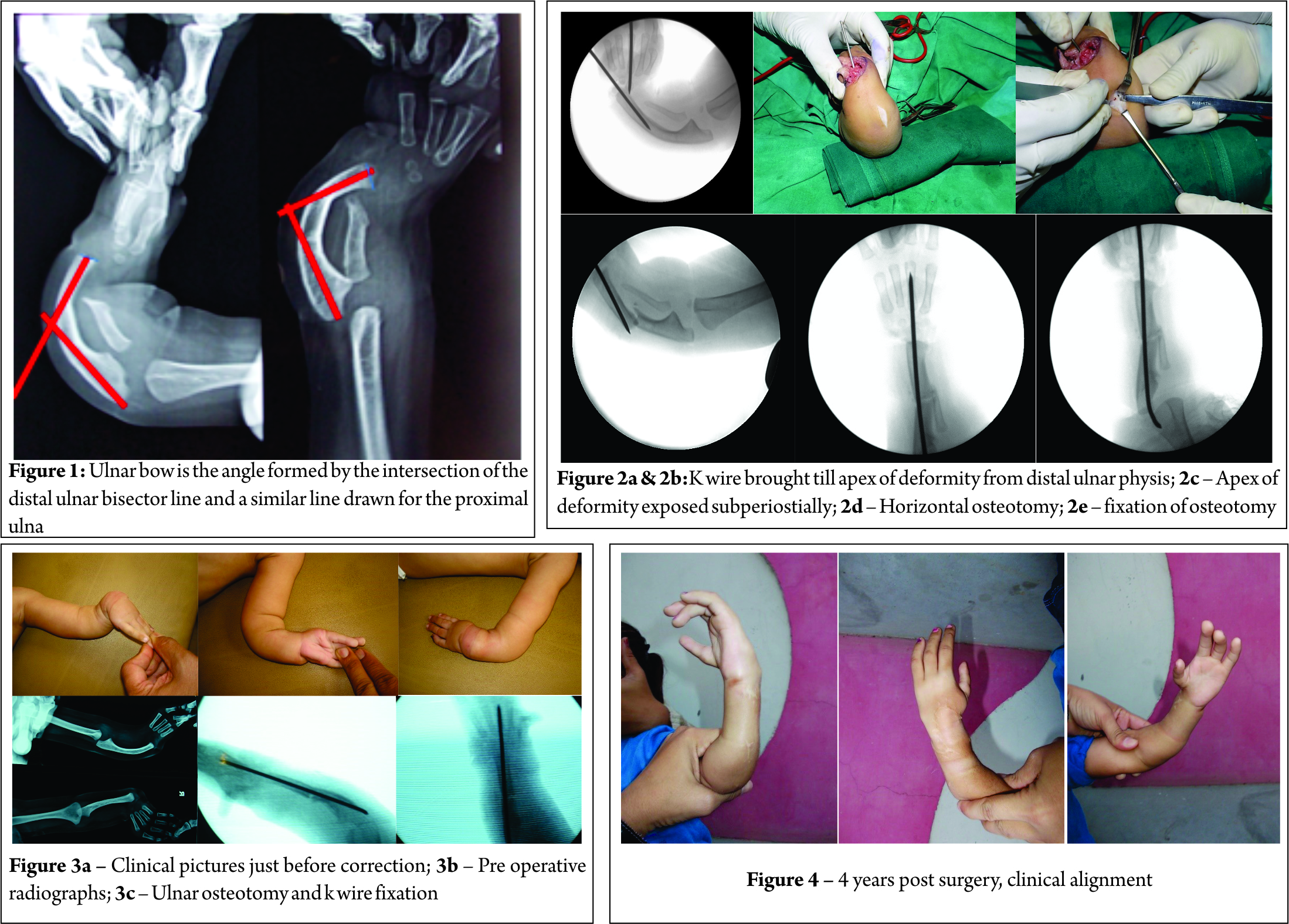
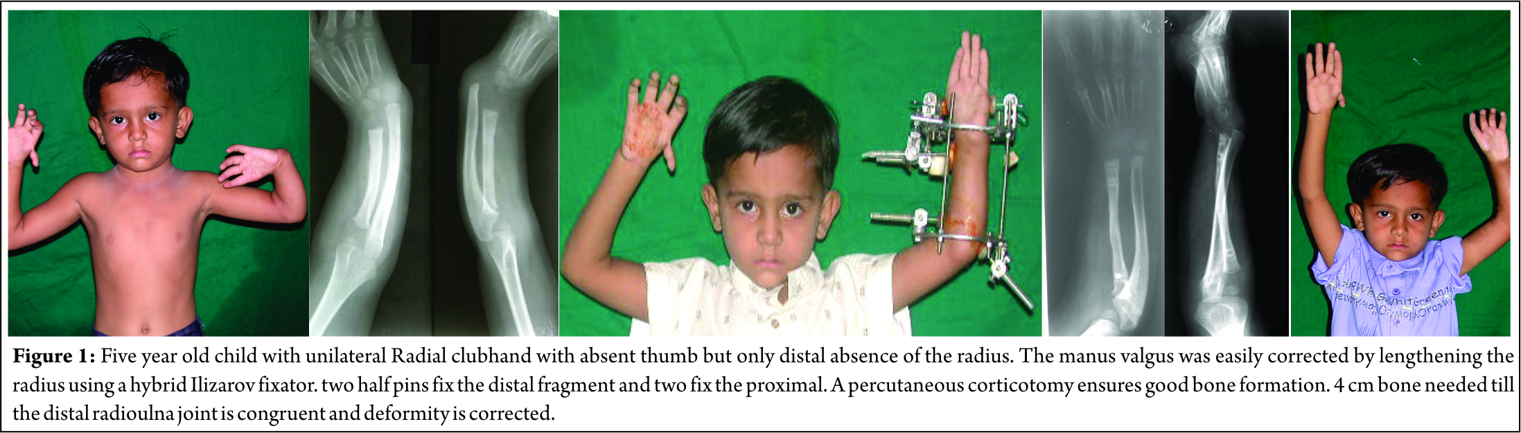
The condition occasionally presents itself as a shortening of the Radius. Distraction lengthening of the distal radius equalizes length to that of the ulna to match it at the distal radial ulnar joint [3,4]. Though uncommon, it is the simplest of all treatment modalities(Fig1).

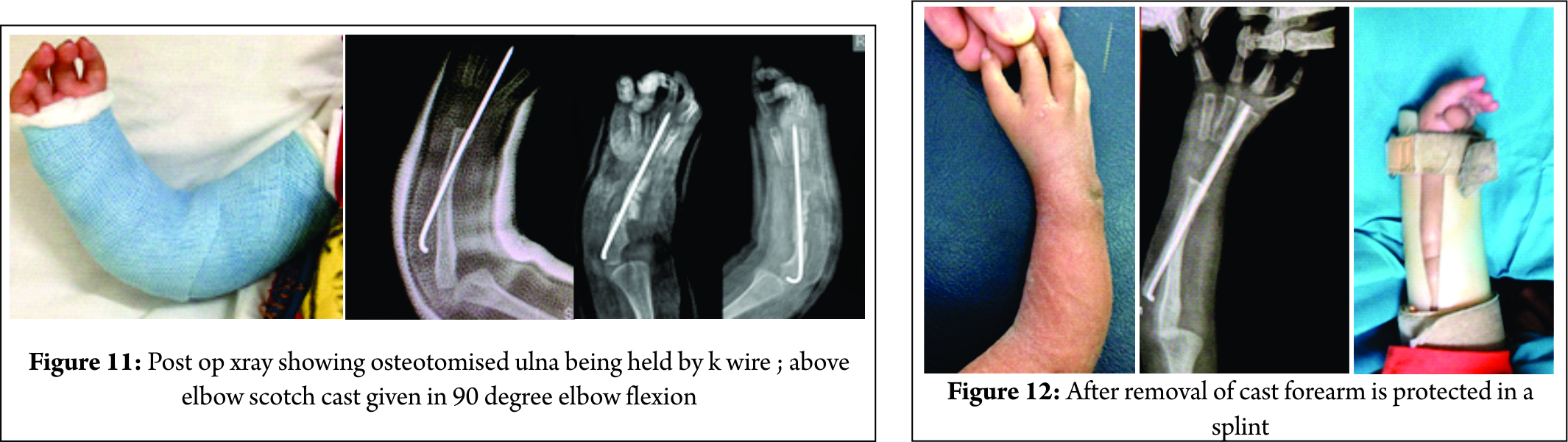
A monolateral fixator is easy to use with two half pins in the proximal and distal fragment each. An angulation translation osteotomy corrects the bowing deformity of the proximal ulna. A large deformity can be corrected percutaneously. In this situation, it is better to perform the surgery using the Ilizarov external fixator. A 5/8 th ring fixed proximally at the elbow is kept open anteriorly to allow flexion of the elbow joint. The distal ulnar ring can be a full one.
The hand is fixed with a ring with wires and half pins. Distraction in the concavity corrects the radial deviation deformity. The hand ring also prevents deformities that may arise with distraction.
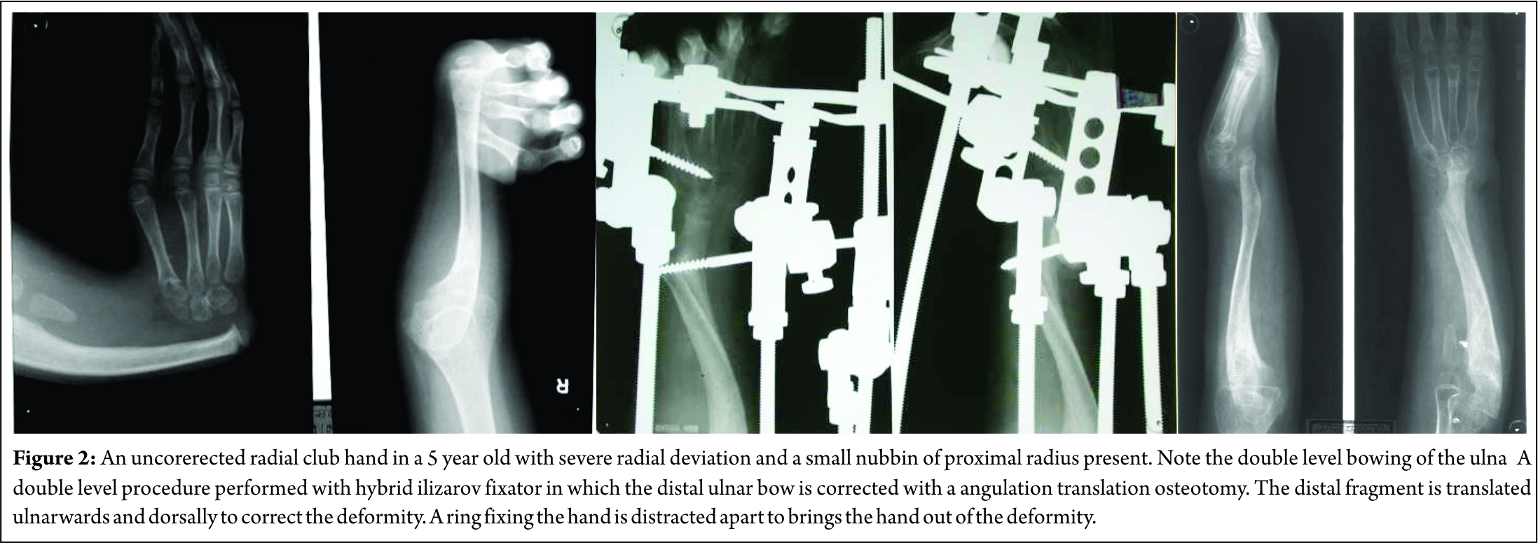
Deformity at the carpus can be corrected with an angulation-translation osteotomy of the distal ulna without resorting to open surgery. Ulnar bow is corrected by angulating the distal ulnar fragment with medial translation. This helps buttress the wrist and improve the appearance of the hand (Fig. 2).


Lengthening of the radius equalizes radio –ulnar length in acquired clubhand due to growth arrest. The aim is to correct the length deficit as well as angular deformity of the lower end of radius and to try and match it to the distal ulna [5,6] . The physeal arrest must also be addressed on its merits and a physeal bar resection must be performed.

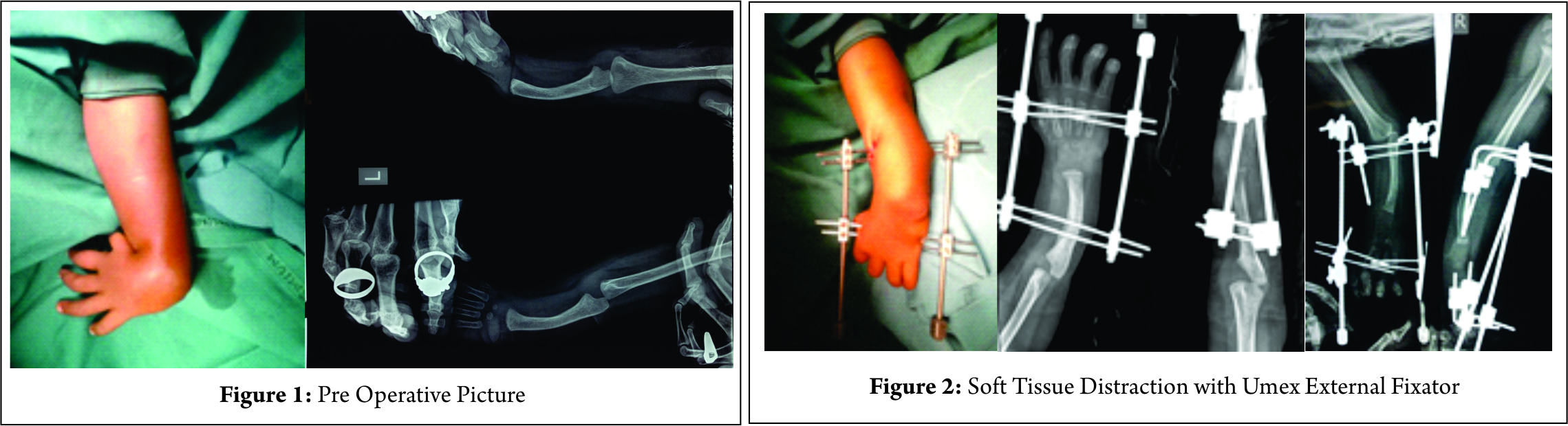
The use of distraction techniques as a precursor to Centralization
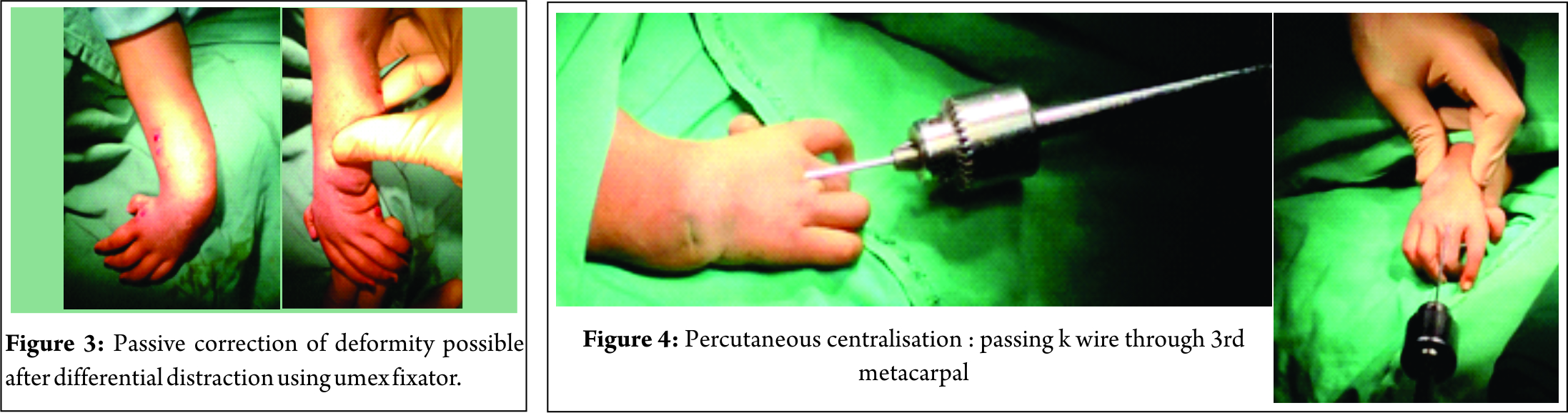
Deformity correction by centralization is popular and the aim is to get the ulna to be collinear with the lunate, capitate and third metacarpal. Extensive soft tissue release with resection of the capsule is needed to get the carpus in line with the ulna. The deficiency of the soft tissues as well as skin on the radial side makes this a difficult task. Many have also described the role of pre-centralization distraction. It is an attractive concept as it may help reduce the extensive soft tissue dissection needed to get the carpus on top of the ulna. The soft tissue deficit causes radial deviation and ulnar subluxation of the wrist. Gradual distraction of the soft tissues doesn’t merely stretch them but lengthens them according to the law of tension stress [7]. A monolateral or circular external fixator is applied to the hand and ulna. Soft tissue elongation allows the hand to be distracted out of radial deviation & volar subluxation to enable the third metacarpal to become collinear with the ulna [ 8,9,10,11,12]. The external fixator retains the lengthened position till soft tissues mature & prevents reshortening. This may reduce the extent of soft tissue dissection needed to achieve correction.
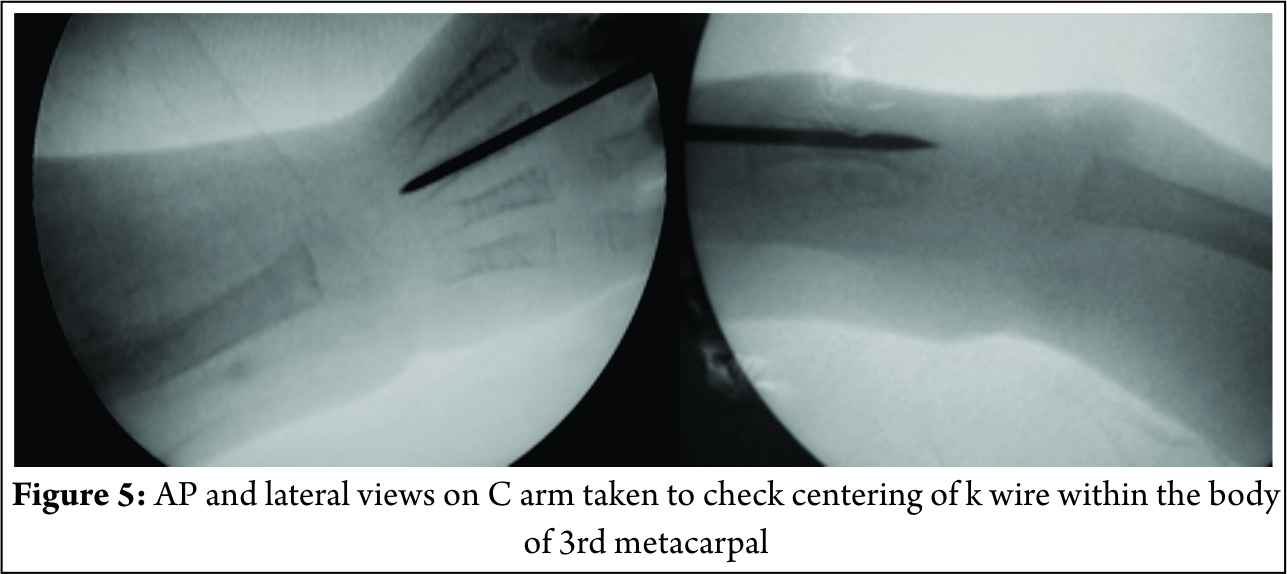
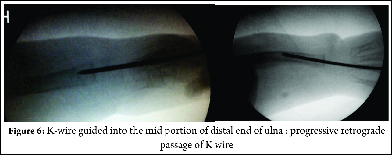
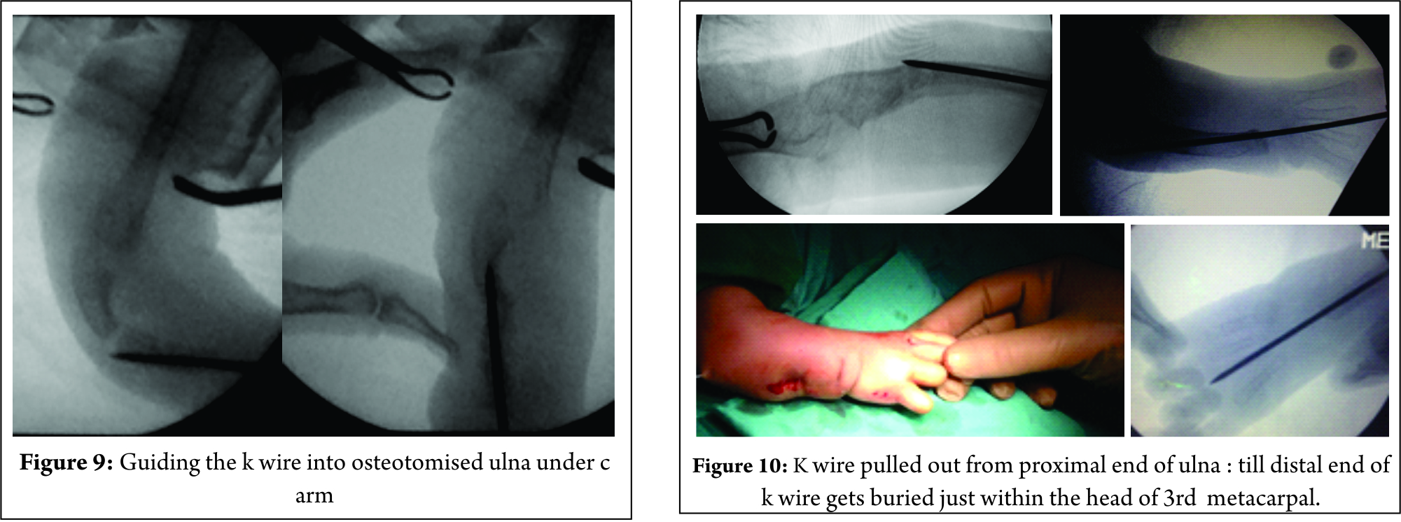
A K wire travels from the third metacarpal going down into the capitate, lunate and ulna. Some prefer to remove the K wire at 12 weeks and retain the correction by either tendon transfers or a brace. Some prefer to leave the K wire in situ. The K wire may be passed without making any preparatory changes in distal ulna. External fixation corrects the deformity maintains the position thereafter. The distal ulna flattens out to match the surfaces of the carpus. Preserved movement and improved appearance gives a good cosmetic and functional result.
The external fixator maintains position and stabilizes the wrist. Some surgeons create a notch in the proximal carpus and insert the distal ulna in it. The notch behaves like a multiplane joint to allow movement and prevents recurrence of deformity.

Distraction techniques as a precursor to Microvascular joint transfer
There is a complete deficit of the radial side of the wrist. Any positioning of the carpus on the distal ulna is therefore unstable. Distracting the carpus out of radial deviation and volar subluxation creates space on the radial side of the wrist. Microvascular technique is used to fill this space with the second metatarsal and metatarsophalangeal joint transfer. The bone is fixed to the ulna in a Y shaped manner [13,14]. The advantage of this method is that a proper joint is created and buttress support to the radial aspect of the wrist is permanent. However, long term studies are lacking on how these transfers perform. The microvascular technique itself is rather complex and such skills may not be available at all centres which makes their application rather limited.

Distraction techniques to correct residual deformities in Radial Clubhand
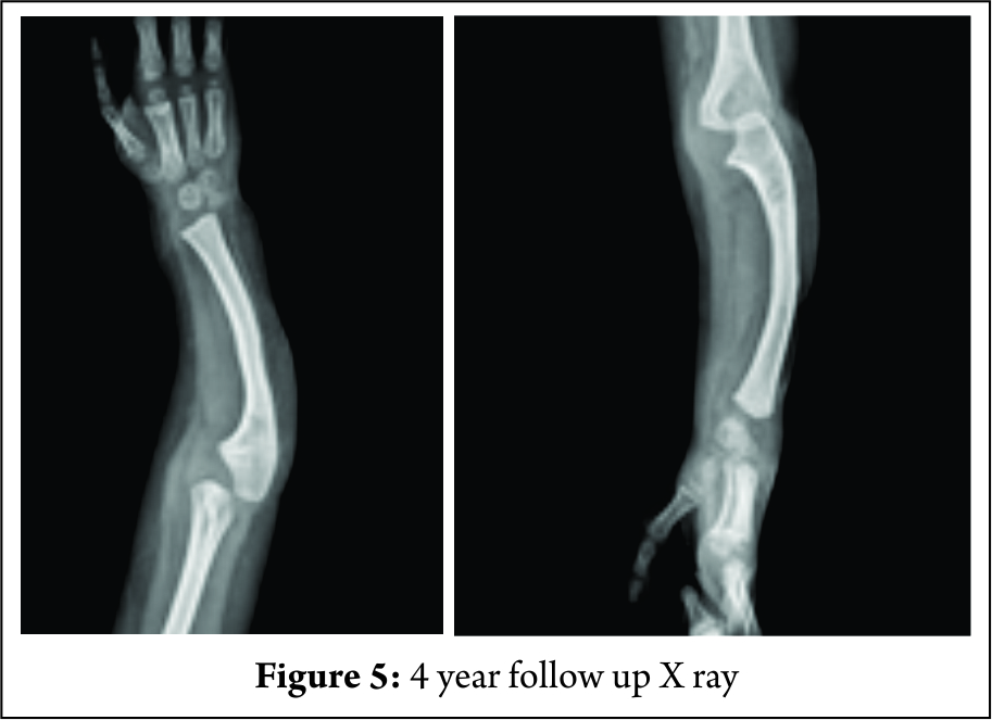
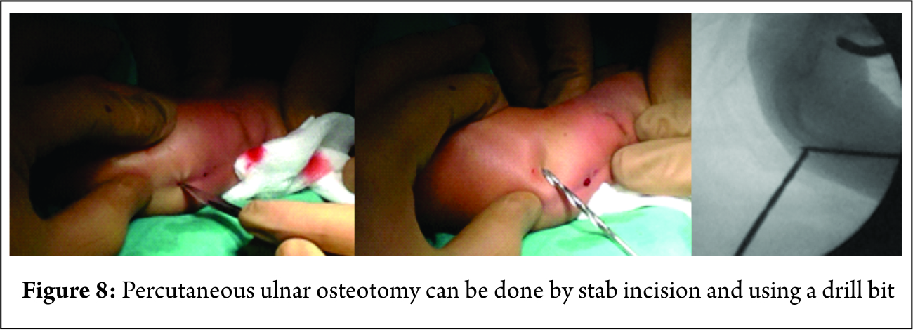
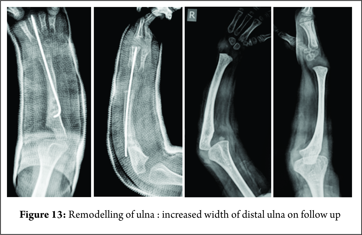
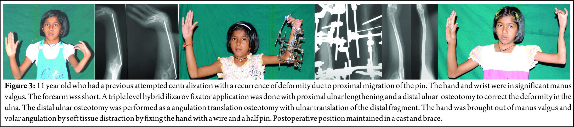
A common complication of centralization is recurrence of the deformity. Since K wires are removed after a few weeks (or may migrate proximally), recurrence of the deformity is likely due to re-shortening of the fibrotic and inelastic soft tissues. The extensive dissection of the distal ulna may damage its blood supply and result in distal ulnar growth arrest with deformities and shortening. In these situations, a circular external fixator [15,16,17] can easily correct the residual deformity by soft tissue distraction of the hand and wrist. The hand is brought out of the radial deviation and volar subluxation and repositioned on top of the distal ulna. Passing a K wire, or creating a trough for the ulna or by a wrist arthrodesis creates stability. Prolonged bracing may also help. Percutaneous osteotomy helps correct deformities at any level in the ulna. Proximal ulnar lengthening can be added as well. (Fig. 3)
Corticotomy lengthening and gradual correction of the angular deformity are possible in the proximal ulna. Compression between the distal ulna and carpus helps fuse the wrist.

Distraction as an adjunct to the operation of Ulnarization
Wrist and finger movement are more important than maintenance of hand forearm angle for long term hand function [18]. However, fingers are usually stiff to start with and cannot be influenced by surgery. Centralization or radialization reduce movement at the wrist even without fusion. Recurrence of the radial deviation and volar subluxation deformity of the wrist is common.
Creating a notch in the wrist with residual deformity may eventually need an arthrodesis. Combined with the restricted movements in the elbow and PIP and DIP joints of the fingers stiffness of the wrist can contribute to functional disability.
Paley described Ulnarization to correct the deformity, retain movement at the wrist and prevent its recurrence [19]. This is an advanced technique of reconstruction of the deformities of radial clubhand. It prevents the problems associated with the previous methods of treatment, namely: recurrence of the deformity, stiffness of the wrist and distal ulnar growth arrest.
Ulnarization shifts the wrist and hand from radial to the ulnar side of the distal ulna. The ulnar border of the distal ulna mechanically prevents radial deviation of the wrist. (Fig. 4).


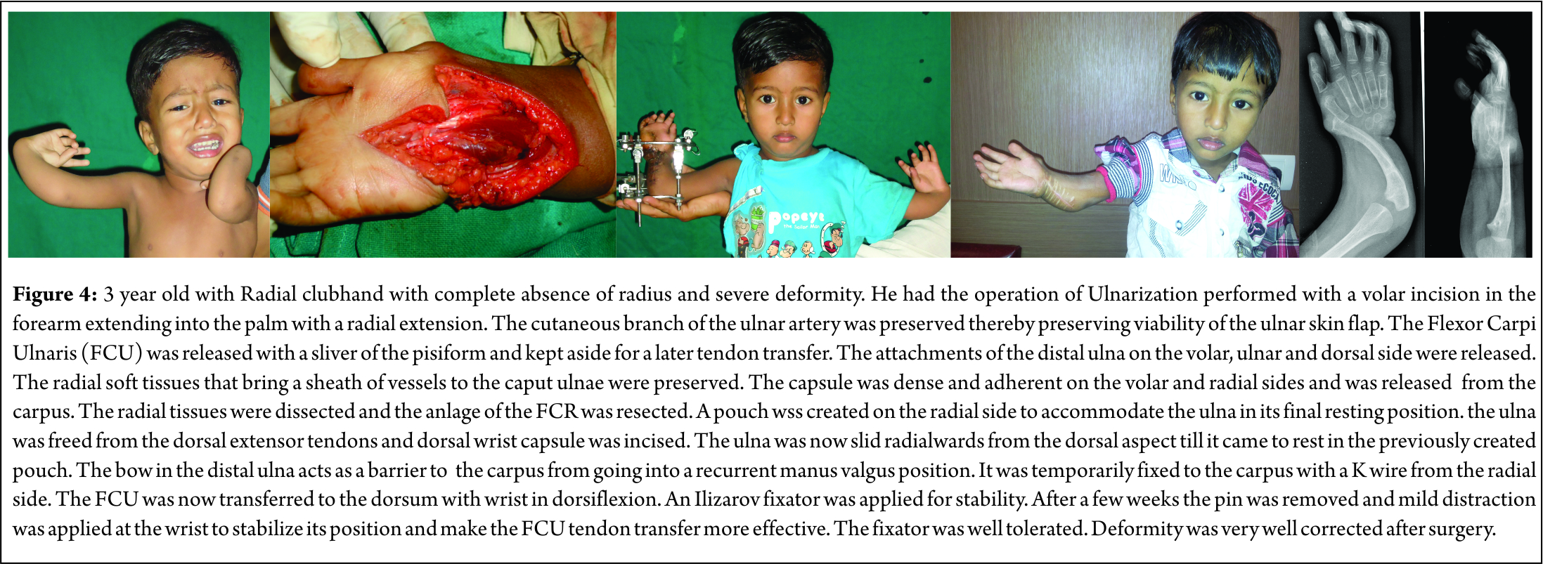
The second part of the operation transfers the flexor carpi ulnaris tendon from the pisiform to the dorsal side of the wrist. The operation can be performed as early as 15-18 months of age. Magnification with a surgical loupe helps preserve small vessels in surgery in very young children. Pollicization of the index finger can be performed at a later date.
The incision begins at ulnar border of lower humerus, extends across the forearm, goes towards radial aspect wrist and then back along the wrist crease in a Z to open in the palm. The radial extension helps to create a pocket on the radial side of the distal forearm and wrist joint in which the distal ulna can be translocated. The blood supply to the distal ulnar epiphysis comes from the radial side and should be preserved while freeing the distal ulna from the wrist capsule. The tendons dorsal to ulna are released by sharp dissection from the distal ulna. Complete release of capsule from the radial, volar and dorsal sides helps mobilize the distal ulna.
Gentle dissection creates a pocket on the radial side of the distal forearm allowing the distal ulna to slide from the dorsum of the wrist towards its radial aspect. Care is taken to prevent subluxation of wrist and hand on the volar or dorsal side of distal ulna. It is fixed to the hand and wrist with a K-wire for a few weeks. Ilizarov fixator fine tunes position of the hand and wrist on the distal ulna. It may also be used for an osteotomy of the proximal ulna if grossly deformed. The distal ring is distracted to improve the tension in the soft tissues and transferred tendons.
Prominence of the distal ulna on the radial side of the wrist looks like a prominent radial styloid. The appearance of the entire forearm and hand is dramatically improved. The FCU is transferred to the dorsal ulnar side of the wrist to the base of the fifth metacarpal. The wrist can dorsiflex due to transferred action of flexor carpi ulnaris. Muscles on the radial side of the wrist are usually absent and unavailable for tendon transfers.
The author has performed five procedures in four patients over the last seven years. Age has ranged from 2 to 18 years of age. Follow-up has now ranged from a period of three years to eight years. A short period of bracing or casting was needed in two of our cases. The improved appearance of the hand was satisfactory for all of our patients. There was a mild recurrence of the volar flexion deformity at the wrist in 2 of five patients. There was very little recurrence of radial deviation deformity. Three had aplasia of the thumb for which they have not yet come for pollicisation. Poor hand function has been chiefly due to lack of the thumb and stiffness of the fingers.

Conclusion

Distraction techniques using monolateral and Ilizarov external fixators have an important role in the treatment of Congenital Radial club hand. They help lengthen the short radius and the shortened ulna along with deformity correction of the ulna either at the proximal or distal level. They ease the operations of centralization of the wrist by reducing the need for extensive soft tissue distraction. Recurrent deformities are easily corrected by distraction techniques. It aids displacement of the carpus to the ulnar border of distal ulna in Ulnarization.  This procedure improves appearance of the hand by correcting the deformity, prevents its recurrence but preserves the mobility of the wrist.


References 

1. Lamb DW. Radial club hand. A continuing study of sixty-eight patients with one hundred and seventeen club hands. J Bone Joint Surg Am. 1977 Jan;59(1):1-13.
2. Bora FW Jr, Osterman AL, Kaneda RR, Esterhai J. Radial club-hand deformity. Long-term follow-up. J Bone Joint Surg Am. 1981 Jun;63(5):741-5.
3. Tetsworth K, Krome J, Paley D. Lengthening and deformity correction of the upper extremity by the Ilizarov technique. Orthop Clin North Am. 1991;22: 689-713.
4. Takagi T, Seki A, Mochida J, Takayama S. Bone lengthening of the radius with temporary external fixation of the wrist for mild radial club hand. J Plast Reconstr Aesthet Surg. 2014 Dec;67(12):1688-93.
5. Zhang X, Duan L, Li Z, Chen X. Callus distraction for the treatment of acquired radial club-hand deformity after osteomyelitis. J Bone Joint Surg Br. 2007 Nov;89(11):1515-8
6. Hosny GA, Kandel WA. Treatment of posttraumatic radial club hand with distraction lengthening. Ann Plast Surg. 2013 Nov;71(5):489-92.
7. Ilizarov G.A. Clinical effect of the tension stress effect for limb lengthening. Clin. Orthop. Rel. Res.1990 Jan (250) 8: 26.
8. Sabharwal S, Finuoli AL, Ghobadi F. Pre-centralization soft tissue distraction for Bayne type IV congenital radial de¬ficiency in children. J Pediatr Orthop 2005;25(3):377-81.
9. Kanojia RK, Sharma N, Kapoor SK. Preliminary soft tissue distraction using external fixator in radial club hand. J Hand Surg Eur Vol. 2008 Oct;33(5):622-7.
10. Thirkannad SM, Burgess RC. A technique for using the Ilizarov fixator for primary centralization in radial clubhand. Tech Hand Up Extrem Surg. 2008 Jun;12(2):71-8.
11. Saini N, Patni P, Gupta S, Chaudhary L, Sharma V. Management of radial clubhand with gradual distraction followed by centralization. Indian J Orthop. 2009 Jul;43(3):292-300.
12. Bhat SB, Kamath AF, Sehgal K, Horn BD, Hosalkar HS. Multi-axial correction system in the treatment of radial club hand. J Child Orthop. 2009 Dec;3(6):493-8.
13. Vilkki SK. Distraction and microvascular epiphysis transfer for radial club hand. J Hand Surg Br. 1998 Aug;23(4):445-52.
14. de Jong JP, Moran SL, Vilkki SK. Changing paradigms in the treatment of radial club hand: microvascular joint transfer for correction of radial deviation and preservation of long-term growth. Clin Orthop Surg. 2012 Mar;4(1):36-44.
15. Kawabata H, Shibata T, Masatomi T, Yasui N. Residual deformity in congenital radial club hands after previous centralisation of the wrist. Ulnar lengthening and correction by the Ilizarov method. J Bone Joint Surg Br. 1998 Sep;80(5):762-5.
16. Damore E, Kozin SH, Thoder JJ, Porter S. The recurrence of deformity after surgical centralization for radial clubhand. J Hand Surg Am. 2000 Jul;25(4):745-51.
17. Shariatzadeh H, Jafari D, Taheri H, Mazhar FN. Recurrence rate after radial club hand surgery in long term follow up. J Res Med Sci. 2009 May;14(3):179-86.
18. Hand function in children with radial longitudinal deficiency Anna Gerber Ekblom, Lars B Dahlin, Hans-Eric Rosberg, Monica Wiig, Michael Werner, Marianne Arner BMC Musculoskeletal Disorders 2013, 14:116.
19. Paley D, Robbins CA. Ulnarization for treatment of radial club hand. Limb Lengthening & Reconstruction Surgery Case Atlas. Switzerland, Springer International 2015 Jan:1-11.


How to Cite this Article: Chaudhary M. The use of distraction techniques in treating radial clubhand. International Journal of Paediatric Orthopaedics Sep-Dec 2016;2(3):12-15.


(Abstract)      (Full Text HTML)      (Download PDF)


Ulna Osteotomy Role – Methods, Timing, Combo Procedures, Recurrences and Re- Osteotomy

Volume 2 | Issue 3 | Sep-Dec 2016 | Page 16-18| Premal Naik, Hitesh Chauhan


Authors : Premal Naik [1], Hitesh Chauhan [1]

[1] Rainbow Rainbow Super Speciality and Children Orthopaedic Hospital, Ahmedabad.

Address of Correspondence
Dr Premal Naik
Rainbow Rainbow Super Speciality and Children Orthopaedic Hospital, Ahmedabad.
Email: premalnaik@gmail.com


Abstract

Ulna bowing is common occurrence in radial club hand and depends on the severity of the deformity. Centralisation takes care of the wrist stability and deformity however ulna deformity if ignored may continue to progress and cause significant forearm deformity. Current recommendation it to perform Ulna osteotomy at the time of index procedure of centralisation, if the ulna deformity is more than 30 degrees. Current article describes the technique and methods of ulna osteotomy.
Keywords: Ulna osteotomy, radial club hand.


Introduction

Congenital radius deficiency, or radial hemimelia, is characterized by a hypoplasia or complete absence of the radius. In radial hemimelia, ulnar bowing plays a significant role in overall deformity. Severity of ulnar bowing is mostly proportional to severity of radial hypoplasia.
Centralization for correction of radial hemimelia was proposed in 1894 [8] and has been modified later on by many surgeons [1, 2, 6, 7, 10]. It has shown significant improvement in overall wrist function and strength but correction of forearm deformity i.e. correction of ulnar bow has not been given due importance.
Progressive ulnar bowing is an important late complication after centralization surgery, when forearm deformity is severe and is not corrected [1, 5, 9]. There remains a dilemma whether to correct ulna bowing during the index procedure or to perform it at a second stage.

Ulnar bowing – Current scenario
Ulnar bow is the angle formed by the intersection of the distal ulnar bisector line and a similar line drawn for the proximal ulna (Fig.1) [3]. Severity of forearm deformity depends on the severity of radial hypoplasia. Radial hemimelia with complete absence of radius (Bayne and Klug type III & IV) present with severe forearm deformity as compared to mild hypoplasia (Bayne and Klug type II).
According to Bayne and Klug and few other reports, ulnar bow was considered to be significant if angular deformity is more than 30°. They did not recommend corrective osteotomy of the ulna if the angular deformity was less than 30° [1] .
According to Geck MJ et.al ulnar osteotomy was performed for ulnar bow greater than 30° and for milder deformity of less than 30° it was done only if needed, to pass the transfixing pin[3].
Timing and method of ulna osteotomy is not defined clearly in literature and depends on surgeon’s preference. It is either performed during index procedure or can be done at a second stage[3, 9]. Few surgeons prefer to correct ulnar angulation at the time of lengthening[4].
In Geck MJ et.al series, 13 ulnar osteotomies were performed along with index procedure and 4 osteotomies were done as secondary procedure. The ulnar osteotomy was performed at the apex of deformity when deformity is more than 30° and in less severe deformity, it was performed wherever k wire could not be passed thorough ulnar shaft. Osteotomy was transfixed with same k wire, which is fixing the wrist. Wire was removed at 8 – 12 weeks. They noted no statistically significant difference of results between osteotomy done along with index procedure or as secondary procedure at the final follow-up. They could achieve statistically significant correction in ulnar bow from preoperative measurement to final follow-up measurement and found that Ulnar osteotomy did not have a deleterious effect on the correction of the wrist deformity [3].
H. Kawabata and colleagues recommended correction of congenital radial club hand by staged procedures. The first is centralization followed by lengthening of the ulna and correction of the angular deformity using the Ilizarov method. In their series mean angular deformity was 42°. Full correction of angular deformity was done in six out of seven patients but at final follow up mean correction was 57 % of initial correction [4].
Deformity recurrence and revision is an important issue in surgical correction of congenital radius deficiency. Revision surgery is mostly attributed to recurrent increased hand forearm angle or increased radial translation at wrist as compared to recurrence of ulnar bow[4, 9].
Due to poor growth potential of ulnar physis, recurrence of forearm deformity after correction is mostly not significant[4]. Geck MJ showed that there was no statistically significant difference between the postoperative and final follow up ulnar bow in patients with and without ulnar osteotomy[8].
According to H. Kawabata, recurrence of ulnar bowing was due to muscle imbalance which was exaggerated by the lengthening. The recurrence was least in a wrist with well-balanced muscle forces. So they proposed first centralization procedure is of great importance for further Illizarov correction[4] .

Authors preferred treatment
We routinely take ‘All In One’ approach for correction of radial club hand. This includes single stage, centralization with tendon transfer and Ulna Osteotomy. Addition of ulnar osteotomy adds very little time and obviates the need for second surgery. In our center we have operated 45 extremities in 40 patients till May 2016. We presented our experience of 24 extremities in 20 patients earlier (POSICON 2013). We could achieve statistically significant improvement in wrist forearm angle and ulnar angulation in all patients. Ulnar angulation was corrected from an average of 380 preoperatively to 130 postoperatively (p value – < 0.0001) and wrist forearm angle was corrected from an average of 410 preoperatively to 130 postoperatively (p value – < 0.0001). We did not have any significant problem related to ulnar osteotomy. We found ulnar osteotomy a useful adjunct in the treatment of radial hemimelia.

Surgical technique
We use either Ewan bilobed flap of lazy S incision. After exposing the wrist a provisional track is made with k wire from distal ulnar epiphysis till apex of deformity (Fig 2a, b). One K wire is then passed from 3rd MC head transfixing carpus over distal ulna and advanced in distal ulnar shaft till apex of deformity (in previously made tract).
Nail tip (at the apex of the deformity) is confirmed under image intensifier guidance. Apex of bow exposed subperiosteally (Fib 2 c) and horizontal osteotomy is done (Fig 2 d), k wire is then advanced in proximal fragment under vision and brought out through tip of olecranon (Fig 2 e). In severe deformities, minimal shortening is done to correct the deformity to avoid excessive stretch and injury to neurovascular bundle. After fixation of wrist and ulna osteotomy, tendon transfer is performed. AE cast is given for 6 weeks, followed by strict splinting. K wire is kept for at least 6 months post operatively. Fig. 2 describes the surgical technique
Illustrated Case: Two month old male child, presented with left radial hemimelia(Fig 3 a). On radiological evaluatoin, there was complete absence of radius with gross bowing of ulnawith pre operatively ulnar angulation of 400 (Fig 3 b) . He underwent ‘All In One correction’ at age of 10 months (Fig 3 c). Child under went pollicisation 1 year after primary surgery. After 4 year child is having good hand function and very good overall wrist and forearm allignment (Fig – 4). On follow up, ulnar angulation was 150 (Fig. – 5).

Conclusion

We have found ulnar osteotomy (along with centralisation and tendon transfer) a very useful and powerful tool in managing radial hemimelia. Ulnar osteotomy adds extra 20-300 correction in a significantly deformed upper limb.
Ulnar osteotomy is a simple procedure and does not add significant extra surgical time. We did not have any significant complications related ulnar osteotomy. We recommend ulnar ostetomy in all cases when angulaton is > 300 or when k wire can not be passed straight through the ulna.


References 

1. Bayne LG, Klug MS. Long-term review of the surgical treatment of radial deficiencies. The Journal of hand surgery. 1987;12:169-179.
2. Buck-Gramcko D. Radialization as a new treatment for radial club hand. The Journal of hand surgery. 1985;10:964-968.
3. Geck MJ, Dorey F, Lawrence JF, Johnson MK. Congenital radius deficiency: radiographic outcome and survivorship analysis. The Journal of hand surgery. 1999;24:1132-1144.
4. Kawabata H, Shibata T, Masatomi T, Yasui N. Residual deformity in congenital radial club hands after previous centralisation of the wrist. Bone & Joint Journal. 1998;80:762-765.
5. Lourie GM, Lins RE. Radial longitudinal deficiency. A review and update. Hand clinics. 1998;14:85-99.
6. Manske PR, McCarroll HR, Swanson K. Centralization of the radial club hand: an ulnar surgical approach. The Journal of hand surgery. 1981;6:423-433.
7. Riordan D. Congenital Absence Of The Radius-a 15-year Follow-up. In: Journal Of Bone And Joint Surgery-american Volume. Journal Bone Joint Surgery Inc 20 Pickering St, Needham, Ma 02192: 1963:1783-1783.
8. Sayre RH. A contribution to the study of club-hand. Trans Am Orthop Assoc. 1894;1:208-216.
9. Shariatzadeh H, Jafari D, Taheri H, Mazhar FN. Recurrence rate after radial club hand surgery in long term follow up. J Res Med Sci. 2009;14:179-186.
10.. Watson HK, Beebe RD, Cruz NI. A centralization procedure for radial clubhand. The Journal of hand surgery. 1984;9:541-547.


How to Cite this Article: Naik P, Chauhan H. Ulna Osteotomy Role – Methods, timing, combo procedures, recurrences and re- osteotomy. International Journal of Paediatric Orthopaedics Sep-Dec 2016;2(3):16-18.


(Abstract)      (Full Text HTML)      (Download PDF)


Percutaneous centralization for Radial Club Hand – A Technical Note

Volume 2 | Issue 3 | Sep-Dec 2016 | Page 19-23 | Sandeep Patwardhan, Kunal Aneja, Ashok Shyam


Authors : Sandeep Patwardhan [1] ,  Dr Kunal Aneja [1], Ashok Shyam [1]

[1]  Sancheti Instittue for orthopaedics and Rehabilitation, Pune, India.
[2] Indian Orthopaedic Research Group, Thane, India.

Address of Correspondence
Dr Sandeep Patwardhan
Sancheti Instittue for orthopaedics and Rehabilitation, Pune, India
Email: sandappa@gmail.com


Abstract

The treatment for radial club hand (Heikel Type III and IV) in stages of soft tissue distraction, followed by centralization of carpus over ulna and later pollicisation has been reported. Traditionally the described procedure for centralization of the carpus over the ulna has been by an open approach, involving placing the growing inferior end of ulna over center of carpus and maintaining it with a K wire. Open centralization procedure is fraught with dangers of damage to cartilage, scarring and stiffness of wrist joint, thus increasing morbidity of the procedure. We describe a new technique of percutaneous closed centralization, taking advantage of pliability of soft tissues achieved by distraction method, thus preserving the biology and function. This technique, to our knowledge, has not been reported earlier and we have found it to be a simple and effective method to achieve centralization for patients with radial club hand.
Keywords: Radial club hand, percutaneous centralisation, ulna osteotomy.


Introduction

Radial club hand is a relatively rare deformity with an incidence of 0.5 per 10,000 live births [1,2,3]. Petit was the first individual to record the first case of radial club hand in the year 1733[4]. Radial club hand is divided by Heikel[5] into four types out of which Type III and IV are considered to be the most common forms[5]. These cases also tend to be associated with the greatest amount of radial deviation of the wrist. Over the years, the management of radial club hand has undergone significant changes. In severe cases (Heikel[5] Type III and IV), requiring surgical intervention for deformity correction and improvement of function, historically various surgical treatment modalities have been performed like ulnar osteotomy and splitting the distal ulna for insertion of the carpus [6], serial casting and open centralisation[7] , ilizarov correction[8,9], reconstruction by a bone graft and non-vascularized epiphyseal transfer[10],vascularized proximal fibular epiphyseal transfer[11]and pollicization[12,13].We believe, a staged treatment of radial club hand involving soft tissue differential distraction, percutaneous centralization and pollicization offers a biological solution to this complex problem, for deformity correction and to enhance function.

Procedure : Staged surgical correction of radial club hand deformity is done in the following manner-

Stage 1 : Soft tissue differential distraction
Done by Using a Universal Mini External Fixator (UMEX fixator) to slowly distract the soft tissues and correct the radial deviation. (Fig. 2 a, b)
1. Under suitable anesthesia and under all aseptic precautions, painting and draping of affected upper limb is done.
2. Two K wires of 1.5 mm diameter each are passed at mid metacarpal level, parallel to each other and passing through all the metacarpals in the coronal plane.
3. Two K wires of 2 mm diameter each are passed parallel to each other at the apex of ulnar bowing, again in the coronal plane.
4. These are connected across carpus using universal mini external fixator for soft tissue distraction, such that one connecting rod is towards the concave (radial) and the other towards the convex (ulnar) side of the deformity. It is usually possible to passively accommodate these K wires in same plane. In case it is not possible to do so, additional frame may be constructed so that the distractors are placed in a coplanar fashion. (Fig. 2c)
5. After 2nd post operative day, differential distraction is started which involves distracting at rate of 1mm/day on radial side in 4 graduated turns of 1/4th mm each and 0.5mm/day on ulnar side in 2 graduated turns of 1/4th mm each. This is taught to the child’s parents for them to continue at home.
6. Adequate pin tract care is taken with regular pin tract dressing on alternate days. Weekly X-rays are taken to confirm centralization of carpus over ulna and distraction across carpus.
7. The guided differential distraction is continued for 4 weeks till the radial deviation of hand is obliterated and the hand is visibly angled slightly to ulnar side.
8. After this is achieved, distraction is stopped and the external fixator is kept in static mode for additional one week, to allow for the soft tissues to stretch. This stretching of soft tissues allows for passive correction of deformity.
Thereafter, as a second stage procedure, external fixator is removed and percutaneous centralization of carpus over ulna is done.
On removal of external fixator frame, it is observed that soft tissue pliability achieved by distraction allows the hand to be placed in over corrected position in relation to ulna. (Fig. 3)

Stage 2: Percutaneous centralization:
1. Neutralizing the hand in relation to forearm, such that there is no visible deviation of hand in both coronal and sagittal planes. This involves holding the hand in line with the forearm in antero posterior view and also ensuring that there is no visible volar deviation of wrist in lateral view. (Fig. 3)


2. After flexing the metacarpals to 90 degrees, to make the head of 3rd metacarpal more prominent and moving the base of the proximal phalynx away, a 2mm K wire is loaded on Jacobs chuck T handle and is advanced in retrograde fashion from head of 3rd metacarpal. It is passed through the distal end of 3rd metacarpal, into its body and exited from its proximal end. (Fig. 4) The positioning of K wire within the substance of 3rd metacarpal is important and hence checked under image intensifier in both anteroposterior and lateral views. (Fig. 5)


3. With manual traction and manipulation, K wire is then centralized over mid portion of distal end of ulnar epiphysis and progressed in a retrograde manner, under image intensifier guidance. The correct selection of entry point in ulna is important to maintain the hand in neutral corrected position in relation to the forearm. Checking it under antero-posterior and lateral views of C-arm ensures that the entry point is in the center of distal ulnar epiphysis. (Fig. 6)


4. In some cases, due to bowing of the ulnar shaft, K wire may hit the apex of ulnar deformity and hence may exit more distally (middle third) in the ulnar shaft. In such cases, to allow for the K wire to exit from proximal 1/3rd aspect of ulnar shaft, following methods can be used to straighten the ulna:
Closed plastic deformation of ulna (Fig. 7) : pressure is applied over the apex of the ulnar bow with the thumb and deformity is corrected under image intensifier guidance. K wire is then guided in a retrograde fashion into the straightened ulna. This K wire prevents the deformed ulna from regaining its original contour.

Percutaneous ulnar osteotomy (Fig 8) : If plastic deformation doesn’t help achieving a straightened ulna, ulnar osteotomy may be warranted. For this, a stab incision is taken over the apex of the ulnar bowing and percutaneous ulnar osteotomy is achieved by drilling with a drill bit. K wire is then guided into osteotomised ulna such that the coronal bowing is maximally corrected (Fig. 9)


Ideally the K wire should exit from the proximal third of ulnar shaft, as proximally as possible. In cases, where minimal bowing of ulna is there and apex of ulnar bowing lies in proximal 1/3rd of ulna, neither osteotomy nor plastic deformation is required and the ulna remodels as the patient grows.
5. K wire is then progressed further proximally through the ulna such that it exits from its proximal end, as proximally as possible. A small nick is made over the skin overlying the K wire, to expose the K wire.


6. K wire is pulled out from the proximal end of ulna with a nose plier till its distal end gets completely buried just within the head of the 3rd metacarpal, to allow for free movements of 3rd Metacarpo-phalangeal (MCP) joint which is checked intra operatively. (Fig.10)


7. Excessive length of the k wire is cut off, bending and burying the remaining portion under the skin and closing the skin incisions primarily with ethilon sutures.
8. To immobilize and externally support the fixation, above elbow scotch cast is given in 90 degrees of elbow flexion for a period of 4 weeks. (FIG 11)
9. After a month, cast is removed and removable brace in the form of PVC ulnar gutter splint is given to support the wrist and forearm in neutral position. (FIG 12)


10. Active and passive range of motion exercises are encouraged for the elbow and fingers of the involved hand.
11. The K wire is kept in situ for maintaining the alignment of carpal bones over the ulna, till the distal ulnar epiphysis widens to accommodate the carpal bones. This distal ulnar remodeling is assessed with radiographs obtained on regular 3 monthly follow up visits. (Fig. 13) We believe this biological plasticity of distal ulna is better served by a closed procedure. K wire is retained in situ for an average period of 1.5 to 2 years, after which it is removed. Pollicisation is done 3 months after centralization procedure.
12. For K wire removal, under image intensifier guidance, a small skin incision is taken over the proximal end of ulna, in line with the intraosseous K wire, and blunt dissection is done to expose the proximal end of K-wire. In those cases where the wire becomes buried under bone due to cortical bone covering it, overlying bone is nibbled out and then the K wire is exposed. K wire is then removed by withdrawing it proximally through this incision. After removal, skin is primarily closed with ethilon sutures.

Discussion

The aim of this technical note is to introduce and describe an innovative technique of percutaneous centralization in staged treatment of patients diagnosed with radial club hand. Advantage of this technique is that it being a closed procedure, involves minimal soft tissue dissection, thus reducing the chances of growth plate injury and is easily reproducible after a short learning curve. The current treatment method followed, where centralization is achieved by an open technique, involves extensive soft tissue dissection and has been associated with high rates of physeal injury [14], recurrent radial deviation [15], and wrist stiffness [16]. Iatrogenic physeal injury can hamper the growth potential of the already shortened forearm and can increase the limb length discrepancy [14]. Complications of the technique described here can be infection leading to K Wire loosening and back out or bending of K wire, recurrence of deformity [15] and restricted wrist range of motion after K Wire removal. Since this staged treatment involves usage of an UMEX external fixator for soft tissue distraction prior to centralization, many parents might be apprehensive with the usage of an external fixator for 6 weeks duration, hence reducing the acceptability of this staged procedure. Many authors have described the usage of an external fixator for radial club hand correction previously. On the other hand, in those patients in whom the radial deviation of hand is passively correctable prior to the start of treatment, primary objective, which is to maintain neutral hand forearm position, can be achieved by directly percutaneously centralizing the hand without soft tissue distraction. A long term follow up study of a large sample size is required to establish the efficacy of this technique and document the complications associated with it.
Summary: The technique of percutaneous centralization of carpal bones over radius is recommended as part of a staged procedure, following soft tissue distraction with UMEX fixator. It is an innovative biological approach relying on realignment of soft tissues and remodeling of distal ulnar physis. It has been described here for treatment of Type III and IV radial club hands and for patients with age equal to or less than 2 years at the time of primary surgical intervention. Advantage of this technique is that it involves minimal soft tissue dissection and is easily reproducible after a short learning curve.

Conclusion

Distraction techniques using monolateral and Ilizarov external fixators have an important role in the treatment of Congenital Radial club hand. They help lengthen the short radius and the shortened ulna along with deformity correction of the ulna either at the proximal or distal level. They ease the operations of centralization of the wrist by reducing the need for extensive soft tissue distraction. Recurrent deformities are easily corrected by distraction techniques. It aids displacement of the carpus to the ulnar border of distal ulna in Ulnarization.  This procedure improves appearance of the hand by correcting the deformity, prevents its recurrence but preserves the mobility of the wrist.


References 

1. McCarthy JJ, Kozin SH, Tuohy C, Cheung E, Davidson RS, Noonan K. External fixation and centralization versus external fixation and ulnar osteotomy: the treatment of radial dysplasia using the resolved total angle of deformity. J Pediatr Orthop 2009;29(7):797–803.
2. Saini N, Patni P, Gupta SP, Chaudhury L, Sharma V. Management of radial clubhand with gradual distraction followed by centralization. Indian J Orthop. 2009;43:292–300.
3. Ekblom AG, Laurell T, Arner M. Epidemiology of congenital upper limb anomalies in 562 children born in 1997 to 2007: a total population study from stockholm,sweden.J Hand Surg Am. 2010 Nov;35(11):1742-54.
4. Pardini AG Jr. Radial dysplasia. Clin Orthop Relat Res. 1968 Mar-Apr;57:153-77.
5. HEIKEL HV. Aplasia and hypoplasa of the radius: studies on 64 cases and on epiphyseal transplantation in rabbits with the imitated defect. Acta Orthop Scand Suppl. 1959;39:1-155.
6. Sayre RH. A contribution to the study of Club Hand. Trans Amer Ortho Assn. 1893;6:208–16.
7. M. Farzan. Congenital Radial Club Hand : Results of centralization in 10 cases Acta Medica Iranica,2005 Vol. 43, No. 1
8. Kawabata H, Yasui N, Ariga K, Shirata T. Bone lengthening with the Ilizarov apparatus for congenital club hands. Tech Hand Up Extrem Surg. 1998 Mar;2(1):72-7.
9. Kanojia RK, Sharma N, Kapoor SK. Preliminary soft tissue distraction using external fixator in radial club hand. J Hand Surg Eur Vol. 2008 Oct;33(5):622-7.
10. Albee FH. Formation of radius congenitally absent: condition seven years after implantation of bone graft. Ann Surg. 1928 Jan; 87(1): 105-10.
11. Medrykowski F, Barbary S, Gibert N, Lascombes P, Dautel G. Vascularized proximal fibular epiphyseal transfer: two cases. Orthop Traumatol Surg Res. 2012 Oct;98(6):728-32.
12. Ceulemans L, Degreef I, Debeer P, De Smet L. Outcome of index finger pollicisation. J Hand Microsurg. 2010 Jun;2(1):13-7.
13. Fujiwara M, Nakamura Y, Nishimatsu H, Fukamizu H. Strategic two-stage approach to radial club hand. J Hand Microsurg. 2010;2:33–7.
14. Sestero AM, Van Heest A, Agel J. Ulnar growth patterns in radial longitudinal deficiency. J Hand Surg Am 2006;31(6):960–967.
15. Damore E, Kozin SH, Thoder JJ, Porter S. The recurrence of deformity after surgical centralization for radial clubhand. J Hand Surg [Am]. 2000 Jul; 25(4):745-751.
16. Shariatzadeh H, Jafari D, Taheri H, Mazhar FN. Recurrence rate after radial club hand surgery in long term follow up. J Res Med Sci. 2009; 14(3):179-86.


How to Cite this Article: Patwardhan S, Aneja K, Shyam AK. Percutaneous centralization for Radial Club Hand – a technical note International Journal of Paediatric Orthopaedics Sep-Dec 2016;2(3):19-23.


(Abstract)      (Full Text HTML)      (Download PDF)