Embryology, Pathoanatomy, Clinical Features, Syndromes with Clinical Relevance in Radial Club Hand
Volume 2 | Issue 2 | May-Aug 2016 | Page 9-11|Anirban Chatterjee, Avi Shah, Rujuta Mehta
Authors :Anirban Chatterjee [1], Avi Shah [2], Rujuta Mehta [2]
[1] Medica Superspecialty Hospital, Kolkata, India
[2] Dept of Paediatric Orthopaedics, Bai Jerbai Wadia Hospital for Children, Mumbai, India
Address of Correspondence
Dr Anirban Chatterjee
Medica Superspecialty Hospital, Kolkata. India
Email: anirbanc28@gmail.com
Abstract
Radial club hand is one of the common upper extremity congenital deficiencies involving partial or complete agenesis of the radius. It is associated with a deformed and subluxed wrist with varying degrees of thumb hypoplasia. Associated soft tissue defects and systemic defects are often found. Though the exact etiology is yet to be ascertained, newer research tends to highlight the importance of the apical ectodermal ridge and its proper development is vital to the formation of the normal forearm structure. The pathoanatomy of the forearm involves a short or absent radius with bowed ulna with a wide spectrum of variability in the formation of carpal bones, preaxial muscles and associated neurovascular structures. Certain syndromes are known to be commonly associated with radial club hand and these need to be specifically investigated before proceeding with treatment of the club hand.
Keywords: radial club hand, embryology, associated syndromes
Introduction
The term radial club hand is applied to children born with complete or partial absence of the radius or the preaxial border of the upper extremity. This produces the characteristic radial deviation of the hand (“mannus valgus”), which is similar to a clubfoot deformity and hence the term “radial club hand”. It is also termed as congenital longitudinal deficiency of the radius and is one of the common congenital upper limb birth defects. Its incidence varies from 1 per 30,000 live births to about 1 per 100,000 [6]. It is ten times more common than ulnar deficiencies and bilateral involvement is seen in 38 – 58% of cases.
Embryology
The hand develops as an upper limb bud during the 4th week of gestation as a mesenchymal condensation covered with ectoderm. The mesoderm part gives rise to muscle, tendon, nerve and bone, while the ectoderm differentiates to form skin, hair and nails. These differentiation elements proceed from proximal to distal as the limb bud elongates.
The development of the limb bud progresses along three axis: proximal to distal, anterior to posterior (i.e. radial or ulnar since the fetal upper limb rotates during development) and a ventral to dorsal. Further differentiation within the limb bud is guided by “organizing tissues” viz. apical ectodermal ridge, zone of polarizing activity and the dorsal ectoderm. Associated molecular pathways include SSH (sonic hedgehog), fibroblast growth factors and Wnt proteins [3]. Wnt proteins are a family of highly conserved secreted signaling molecules that regulate cell to cell interaction during development and adult tissue homeostasis. At 33 days of gestation the hand resembles a paddle without individually distinguishable digits. The five rays of metacarpals and phalanges get defined by 41 days. A process of programmed cell death leads to separation of the five rays into four web spaces between 47 days and 54 days. By 7 weeks the mesenchymal condensation that will eventually produce each of the bones of the carpus becomes evident, while the major appendicular bones get defined by 8 weeks. At the end of the embryonic period all future bones are cartilaginous models of their adult form and distal phalangeal tufts begin their ossification [5].
Etiology
In the 19th century, the etiology of radial club hand was theorized to be either a congenital absence or an acquired defect secondary to syphilis. Yet another theory spoke of abnormal pressure upon the embryo along the radial bud between the third and seventh week of gestation in 1895.
No specific etiology has yet been identified though genetic and environmental factors have been implicated. Most cases seen are sporadic in nature with no specific implicating factor 4. Lamb, in 1977, reviewed 117 radial club hands in 68 patients over a 15 year period, including those exposed to thalidomide (26 cases) and concluded that damage to the apical ectoderm on the anterior aspect of the developing limb bud led to the deformity [7]. Most authors who studied embryology of radial hemimelia by consensus are in favor of insult to the apical ectodermal ridge (AER). This structure is a thickened layer of ectoderm that directs differentiation of the underlying mesenchymal tissue and limb formation [2].Therefore, a defect of the AER is the most probable cause of radial club hand, with the extent of deformity related to the degree and extent of the AER absence [3].Removal of a portion of the AER in chick embryos has produced anomalies similar to radial club hand.
Newer evidence has postulated that decreased limb volume with intact SHH expression can be the causative mechanism. This leads to preservation of the posterior (ulnar) elements, while causing an overall decrease in limb length. Researchers have achieved some success in duplicating similar limb deficiencies in animal models by progressively reducing the apical ectoderm ridge and its associated fibroblast growth factors [1, 2]. Different molecular pathways influence cell growth and apoptosis, thus changing the limb volume during the growth and development of the limb bud. Simultaneous development of other organ systems are often affected leading to various associated syndromes like Holt Oram syndrome, TAR syndrome, Fanconi anemia and VACTERL abnormalities.
Patho-anatomy
Four types of radiological radial dysplasia are evident:
1) Short distal radius: radius is present but short distally. The shortening is due to delayed appearance of distal radial epiphysis and its deficient growth. The thumb is hypoplastic and radial carpals may be absent or hypoplastic.
2) Hypoplastic radius: radius is short and ulna proportionately bowed. Both proximal and distal radial epiphyses have growth deficiency
3) Partial absence of radius: radius is partially absent in its distal middle or proximal third. The ulna is short, thickened and bowed. The carpus is unsupported and the hand rolls around the distal end of the ulna. Hand function is usually poor. (Fig. 1)
4) Total absence of radius: most commonly encountered sub type. There is no radius at all, the ulna is bowed and hand function is poor. (Fig. 2)
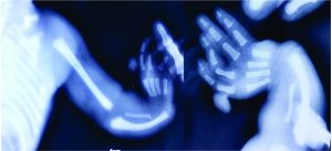
Figure 1 a & b: Radiograph of Type 3 radial club hand showing short radius with bowing of ulna and rudimentary thumb.
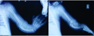
Figure 2: Radiograph of Type 4 club hand showing complete absence of radius with bowing of ulna and rudimentary thumb
Associated abnormalities
a) Local:
Bony: The degree of pre-axial deficiency varies from thumb hypoplasia to complete absence. It may be associated with absence of carpal bones especially the radial ones. The carpal deficiencies are related to the extent of thumb deficiency. For example with an absent thumb the radial carpal bones are absent, while with a complete but hypoplastic thumb, the scaphoid and lunate are hypoplastic. The patterns of thumb hypoplasia are given in Table 2 [6]. Whole or part of the radius may be absent and in severe cases the elbow joint may be poorly formed. The forearm is usually short (50 – 75%) as compared to a normal contralateral forearm [9].
Muscular: these vary in proportion with the skeletal defects. The preaxial muscles (extensors arising from the lateral epicondyle) are frequently deficient. The brachio-radialis, usually present in 50% of cases, inserts into the radial side of the hand and acts as a strong tethering deforming force. The ECRB and ECRL are usually deficient. The thenar muscles are defective in proportion to the thumb deficiency. The post axial muscles (arising from medial epicondyle – pronator teres, FCU, FCR) though well differentiated have abnormal insertions and act as radial deviators of the hand.
Neurovascular: the radial artery is usually absent. The Anterior interooseous artery (arising from the ulnar artery, which is usually normal) supplies the radial part of the forearm. The Ulnar and Median nerves are usually unaffected. The superficial branch of the radial nerve is deficient from the level of the lateral epicondyle. Its function is replaced by a branch of the medial nerve, which runs a course parallel to the absent dorsal cutaneous branch of the superficial radial nerve.
Systemic: In approximately 25% of patients, associated cardiac, gastrointestinal and renal abnormalities may be found. The most frequently associated syndromes include Holt – Oram syndrome, Fanconi anemia, Thrombocytopenia Absent Radius, and VATER (vertebral segmentation deficiencies, anal atresia, Tracheo – esophageal fistula, renal abnormalities and radial ray deficiencies). Trisomy 13 and 18 are also known to be associated with radial deficiencies. In cases with fetal valproate syndrome there is usually an associated syndactyly of the 2nd or 3rd web, which hampers the results of subsequent pollicisation (Fig.. 3).(personal observation of authors )
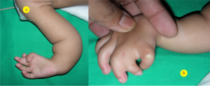
Figure 3 a & b: Clinical picture of child with fetal valproate syndrome showing syndactyly of 2nd web space and hypoplastic thumb and index finger
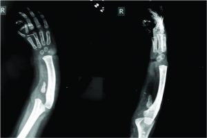
Figure 3 c: Clinical picture of child with fetal valproate syndrome showing syndactyly of 2nd web space and hypoplastic thumb and index finger
In addition to these conditions, a variety of associated musculoskeletal deformities appear sporadically. These include cleft palate, clubfoot, kyphosis, scoliosis, torticollis, and rib deformities. For most of the time in clinical practice the following syndromes are most often encountered by the surgeon and hence are listed in tabular form.
Investigations with relation to syndromes.
Children with VACTERL syndrome warrant additional evaluation for spinal abnormalities, such as congenital scoliosis, and require radiographs of the spinal column. Children with VACTERL syndrome often appear similar to children with Fanconi anemia; they are of small stature, have feeding difficulties, and have similar musculoskeletal anomalies. Therefore, a chromosomal challenge test is warranted in a child with a presumed diagnosis of VACTERL syndrome.
Hematological, The most devastating associated hematological condition is Fanconi anemia. Children with Fanconi anemia do not have signs of bone marrow failure at birth; therefore, the diagnosis is not initially apparent. The majority of children experience signs of aplastic anemia between the ages of 3 and 12 years (median age of 7 years). However, a chromosomal challenge test is available that allows detection of the disease prior to the onset of bone marrow failure. This assay tests a sample of the child’s lymphocytes to diepoxybutane or mitomycin C, which cause chromosomes within Fanconi anemia cells to break and rearrange. In contrast, lymphocytes in unaffected children are stable to these agents.
Because bone marrow transplant is the only cure for Fanconi anemia, this prefatory diagnosis is crucial for the child and family. Early diagnosis provides ample time to search for a suitable bone marrow donor or consider preimplantation genetic diagnosis (PGD). PGD is a sophisticated technique that involves in vitro fertilization, sampling of the blastocytes to ensure human leukocyte antigen (HLA) similarity without Fanconi disease, and re-implantation until birth. At delivery, cord blood is harvested from the newborn and used as a source of stem cell transplant for the affected sibling. Since PGD takes time, early detection via a chromosomal challenge test is critical and may ultimately save the affected child.

Figure 4. A: clinical picture of a child with TAR syndrome and radial club hand b: AP Radiograph showing “carpal height , as measured between the transverse axis of the metacarpal bases and transverse axis of the ulna. Its an important baseline assessment before starting distraction, for early detection of carpal distraction. c: Measurement of lateral axis deviation between the distal end ulna long axis and 3rd MC long axis, to judge the degree of correction required intra operatively
Summary
The management of radial hemimelia is by no means easy, it is a subject which is still unclear as regards to its exact embryology and causation and is a potent field for considerable research. Some day when all the questions are answered we may have a complete understanding of the cause and hence this may lead us to the optimal solution for this varied deformity.
References
1. Mariani F, Ahn C, Martin G. Genetic evidence that FGFs have an instructive role in limb proximal–distal patterning. Nature. 2008; 453(7193):401-405.
2. Sun X, Mariani F, Martin G. Functions of FGF signaling from the apical ectodermal ridge in limb development. Nature. 2002; 418(6897):501-508.
3. Manske P, Oberg K Classification and Developmental Biology of Congenital Anomalies of the Hand and Upper Extremity. J Bone Joint Surgery (Am). 2009; 91(4):3.
4. Kelikian H, Doumanian A, Congenital Anomalies of the Hand Part I. J Bone Joint Surg (Am).1957; 39 (5): 1002 -1019.
5. Light T: Congenital malformations & deformities of the hand. In Instructional Course lectures Vol 38, 1989, AAOS.
6. Conrad M, Ezaki M. Fewer than 10: Oligodactyly-Diagnoses and patterns of malformation. J Am Soc Surg Hand. 2002; 2(3):110-120.
7. Lamb W. Radial club hand. A continuing study of sixty-eight patients with one hundred and seventeen club hands. J Bone Joint Surg Am 1977; 59 (1): 1-13.
8. Saunders W. The proximo-distal sequence of origin of the parts of the chick wing and the role of the ectoderm. 1948. J Exp Zool.1998; 282(6):628-68.
9. Canale, ST and Beaty, JH. (Eds 8) (2007). Campbell’s operative orthopedics. Philadelphia: Mosby Elsevier.
| How to Cite this Article:Chatterjee A, Shah A, Mehta R. Embryology, Pathoanatomy, Clinical Features, Syndromes with Clinical Relevance in Radial Club Hand . International Journal of Paediatric Orthopaedics May-Aug 2016;2(2):9-11. |
