Open reductions of Paediatric Supracondylar Humerus Fractures- When, How and, Risks
Vol 1 | Issue 1 | July-Sep 2015 | page:16-18 | Ashish Ranade, Gauri Oka.
Authors : Ashish Ranade[1], Gauri Oka[1].
[1] Dept. of Orthopaedics, Deenanath Mangeshkar Hospital, Pune 411004.
Address of Correspondence
Dr Ashish Ranade
Dept. of Orthopaedics, Deenanath Mangeshkar Hospital, Pune 411004.
Email address:ashishranade@yahoo.com
Abstract
Supracondylar humerus fracture is one of the commonest fractures in pediatric elbow. Usually closed reduction and percutaneous pinning is the preferred treatment for most of the displaced fractures. Nowadays closed reduction and percutaneous pinning has become standard of care for majority of displaced supracondylar humerus fractures. Rarely, an open reduction via appropriate approach becomes necessary. Various types of approaches that have been described are anterior, posterior, medial, lateral, and combined approaches. There is ambiguity of information as to selection of approach for doing open reduction in a supracondylar humerus fracture. There is debate about timing of treatment, approach selection and indications for doing open reduction.1 In this article we discuss indications, various types of approaches with their pros and cons and risks involved in open reduction of supracondylar humerus fractures in children.
Keywords: Supracondylar humerus fracture, open reduction, surgical approach.
Introduction
Supracondylar humerus fracture (SHF) is one of the commonest fractures in pediatric elbow. Nowadays closed reduction and percutaneous pinning has become standard of care for majority of displaced supracondylar humerus fractures. Rarely, an open reduction via appropriate approach becomes necessary. Various types of approaches that have been described are anterior, posterior, medial, lateral, and combined approaches. There is ambiguity of information as to selection of approach for doing open reduction in a supracondylar humerus fracture. There is debate about timing of treatment, approach selection and indications for doing open reduction [1]. In this article we discuss indications, various types of approaches with their pros and cons and risks involved in open reduction of supracondylar humerus fractures in children.
Case Example
A 9 year old boy was referred for the treatment of left supracondylar humerus fracture. He had sustained an injury following fall from a tree 10 days ago and was put in an above elbow splint in his village. On examination, radial pulse was present and he was neurologically intact.

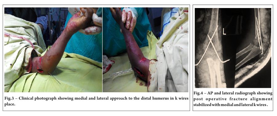
The elbow was grossly swollen and there was deep abrasion with blister formation along the elbow crease on the anterior aspect. (Figure 1) There was ecchymosis along anterior aspect of elbow. The radiographs showed posterolaterally displaced type III supracondylar humerus fracture (Figure 2). Under general anaesthesia, closed reduction was attempted. Satisfactory reduction could not be achieved by closed means. Hence, a decision was made to perform open reduction. Considering the anterior wound, combined medial and lateral approach was chosen. Initially, a medial incision was made and the bony spike of the proximal fragment was separated from the brachialis fibres and the median nerve. At this point, reduction was attempted again. In view of difficulty in getting a satisfactory alignment, a lateral incision was made and the interposing tissues were removed. Periosteum was found to be torn on both sides. (Figure 3)After achieving open reduction, the fracture was fixed with crossed k wires and maintained in an AE slab for 3 weeks(Figure 4). Postoperatively, the patient made uneventful recovery and the fracture healed well in a satisfactory position. The elbow had 5 degrees loss of terminal flexion.
Discussion
Open reduction has been indicated for fractures with vascular injuries, signs of compartment syndrome, failure of closed reduction to achieve satisfactory alignment, and for severe swelling interfering to achieve good reduction [2-7]. In present day scenario, the main indication is failure to achieve satisfactory reduction by closed methods. This could be because of several factors such as instability of the fracture or interposition of neurovascular bundle or brachialis muscle. The overall proportion of supracondylar humerus fractures needing open reduction varies between 3 to 46% based on various studies[2,8-10]. This rate varies between centres and some centres may prefer to do open reductions than using closed methods. Delayed presentation of the fracture is one of the most important factors while discussing open reduction for supracondylar humerus fractures[ 5].
There are several options available for approach selection. There is no clear superiority of one approach over another. Mazzini and co-authors have published a systematic review of literature pertaining to surgical approaches in the treatment of open reduction and pinning[11]. In this review, authors found high frequency of poor results in terms of functional outcomes with posterior approach. High frequency of excellent results was found with the lateral and medial approach and a high frequency of good results within the anterior approach group. A Canadian study sites buttonholing of the proximal fragment through the brachialis muscle and interposition of joint capsule or periosteum between fragments[12]. With the posterior approach , anterior structures such as brachialis, and the neurovascular bundle cannot be accessed and probably the posterior scar leads to limitation of movements of the elbow joint[13]. In the same article, authors have found the change in the carrying angle (cosmetic outcome) as the most common complication seen after an open reduction via the posterior or lateral approach. However, relatively newer studies utilizing posterior approach do not report these complications [7,14]. Medial column communition and internal rotation and/or varus tilt of the distal fragment may be addressed sufficiently through lateral/posterior approach. In review by Mazzini et al, the time to union remains the same irrespective of the approach used. There was higher tendency of ulnar nerve injury in the posterior and lateral approach group. This is attributed to lack of direct visalization of ulnar nerve. Based on the findings, authors recommend anteromedial approach for open reduction[11].
While choosing an approach, one must take into consideration surgeon’s experience and the anatomical structures involved. It is known from various studies that fracture union time and rate of approach related complications are similar with various approaches [7,11].
In a study by Aslan and co-authors, clinical and radiographic results of children with Gartland type 3 supracondylar humerus treated with primary open reduction using four different approaches were studied [7]. Fifty eight patients were treated with either anterior, medial, lateral and , posterior approach. Choice of approach was decided by fracture pattern and neurovascular injury. All fractures were fixed with two lateral entry k wires or crossed k wires as per surgeon’s preference. In this series, three quarters of patients were operated within 24 hours since injury. Flynn criteria were used to measure outcomes. The outcome was comparable in all groups.
Ozkoc and co-authors studied 99 patients with supracondylar humerus fracture. In this group, 44 patients were treated with primary open reduction and k wire fixation and 55 were treated with closed reduction and percutaneous pinning. They found that in the open group the average loss of extension was 6 degrees compared to 0.6 degrees in the closed group[2].
Koudstaal and colleagues have reported the use of anterior approach in 26 children [15]. In another study, Ay and co-authors report their experience of using the anterior approach in 61 children [16]. In both these studies, a transverse incision was used in the antecubital fossa. In both studies, excellent results were noted without any significant loss of elbow movement.
In summary, various options are available for performing an open reduction of a supracondylar humerus fracture. The anterior approach certainly offers advantages of direct visualisation and retraction of entrapped structures. The treating surgeon must choose the appropriate approach based on the indication for open reduction.
Author’s preferred treatment
Our indications for open reduction are as follows:
1) Vascular compromise or disappearance of pulse after doing closed reduction- In this scenario, we suspect the brachial artery likely to be caught between fracture fragments. Hence, we perform an exploration via the anterolateral or anteromedial approach. The vascular structures are explored and reduction of fragments is achieved under vision. We undertake this approach with a vascular/plastic surgeon available in the operation theatre in case the need for vascular repair arises.
2) Inability to achieve satisfactory reduction by closed method- Usually this is encountered in late presentation of fractures with severely swollen elbow. Usually, attempts of closed reduction are made and if satisfactory reduction cannot be achieved, then open reduction is performed. Our preferred approach for this type is usually the anterior approach. However when skin conditions do not permit anterior approach, then a medial and/or lateral approach depending upon the fracture configuration is used.
Open fractures: Usually there is an anterior wound. Anterior approach is used in these cases.
References
1. Mulpuri K, Wilkins K. The treatment of displaced supracondylar humerus fractures: evidence based guideline. J Pediatr Orthop 2012;32:S143-S152
2. Ozkoc G, Gone U, Kayaalp A, Teker K, Peker TT. Displaced supracondylar humeral fractures in children: open reduction vs. closed reduction and pinning. Arch Orthop Trauma Surg 2004; 124:547-551.
3. Cramer KE, Devito DP, Green NE. Comparison of closed reduction and percutaneous pinning versus open reduction and percutaneous pinning in displaced supracondylar fractures of the humerus in children. J Orthop Trauma 1992;6:407-412.
4. Oh CW, Park BC, Kim PT, Park IH, Kyung HS, Ihn JC. Completely displaced supracondylar humerus fractures in children: results of open reduction versus closed reduction. J Orthop Sci 2003;8:137-141
5. Walmsley PJ, Kelly MB, Robb JE, Annan IH, Porter DE. Delay increases the need for open reduction of type –III supracondylar fractures of the humerus. J Bone Joint Surg Br 2006;88:528-530.
6. Mulhall KJ, Abuzakuk T, Curtin W, O;Sullivan M. Displaced supracondylar fractures of the humerus in children. Int Orthop 2000;24:221-223.
7. Aslan A, Konya MN, Ozdemir A, Yougancigil H, Maralcan G, Uysal E. Open reduction and pinning for the treatment of Gartland extension type III supracondylar humeral fractures in children. Strat Trauma Limb Recon 2014;9:79-88.
8. Aktekin CN, Toprak A, Ozturk AM, Altay M, Ozkurt B, Tabak AY. Open reduction via posterior triceps sparing approach in comparison with closed treatment of posteromedial displaced Gartland type III supracondylar humerus fractures. J Pediatr Orthop B 2008;17:171-178.
9. Gupta N, Kay RM, Leitch K, Femino JD, Tolo VT, Skaggs DL. Effect of surgical delay in perioperative complications and need for open reduction in supracondylar humerus fractures in children. J Pediatr Orthop 2004;24:245-248.
10. Reitman RD, Waters P, Millis M. Open reduction and internal fixation for supracondylar humerus fractures in children. J Pediatr Orthop 2001;21:157-161.
11. Mazzini JP, Martin JR, Esteban EMA. Surgical approaches for open reduction and pinning in severely displaced supracondylar humerus fractures in children: a systematic review. J Child Orthop 2010;4:143-152.
12. Fleiriau-Chateau P, McIntyre W, Letts WM. An analysis of open reduction of irreducible supracondylar fractures of the humerus in children. Can J Surg 1998;41(2):112-118.
13. Gruber MA, Hudson OC. Supracondylar fracture of the humerus in childhood. End results study of open reduction. J Bone Joint Surg Am 1964;46:1245-1252.
14. Sibly TF, Briggs PJ, Gibson MJ. Supracondylar fractures of the humerus in childhood: Range of movements following the posterior approach to open reduction. Injury 1991;22(6):456-458.
15. Koudstaal MJ, De Ridder VA, De Lange S, et al. Pediatric supracondylar humerus fractures: The anterior approach. J Orthop Trauma 2002;16(6):409-412.
16. Ay S, Akinnci M, Kamiloglu S, Ercetin O. Open reduction of displaced pediatric supracondylar humeral fracture through the anterior cubital approach. J Pediatr Orthop 2005;25:149-153
.
| How to Cite this Article: Ranade A, Oka G. Open reductions- When, How and, Risks. International Journal of Paediatric Orthopaedics July-Sep 2015;1(1):16-18. |